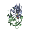
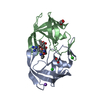
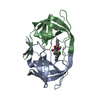
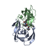
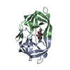
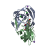
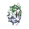
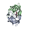
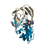
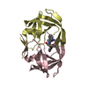
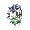
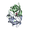
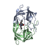
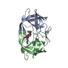
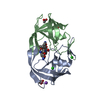
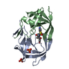
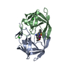
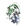
#1: Journal: Bioorg.Med.Chem. / Year: 2003 Title: Design and Synthesis of HIV-1 Protease Inhibitors. Novel Tetrahydrofuran P2 and P2'-Groups Interacting with Asp29 and 30 of the HIV-1 Protease. Determination of Binding from X-Ray Crystal ...Title: Design and Synthesis of HIV-1 Protease Inhibitors. Novel Tetrahydrofuran P2 and P2'-Groups Interacting with Asp29 and 30 of the HIV-1 Protease. Determination of Binding from X-Ray Crystal Structure of Inhibitor Protease Complex Authors: Oscarsson, K. / Lahmann, M. / Lindberg, J. / Kangasmetsae, J. / Unge, T. / Oscarson, S. / Hallberg, A. / Samuelsson, B.
History
Deposition
Nov 2, 2004
Deposition site: PDBE / Processing site: PDBE
Revision 1.0
Nov 4, 2004
Provider: repository / Type: Initial release
Revision 1.1
May 8, 2011
Group: Version format compliance
Revision 1.2
Jul 13, 2011
Group: Version format compliance
Revision 1.3
Jan 17, 2018
Group: Data collection / Category: diffrn_source / Item: _diffrn_source.pdbx_synchrotron_site
SHEET DETERMINATION METHOD: DSSP THE SHEETS PRESENTED AS "AB" IN EACH CHAIN ON SHEET RECORDS BELOW ... SHEET DETERMINATION METHOD: DSSP THE SHEETS PRESENTED AS "AB" IN EACH CHAIN ON SHEET RECORDS BELOW IS ACTUALLY AN 7-STRANDED BARREL THIS IS REPRESENTED BY A 8-STRANDED SHEET IN WHICH THE FIRST AND LAST STRANDS ARE IDENTICAL. THE SHEETS PRESENTED AS "BA" IN EACH CHAIN ON SHEET RECORDS BELOW IS ACTUALLY AN 7-STRANDED BARREL THIS IS REPRESENTED BY A 8-STRANDED SHEET IN WHICH THE FIRST AND LAST STRANDS ARE IDENTICAL.
Method to determine structure: MOLECULAR REPLACEMENT / Resolution: 2→50 Å / Data cutoff high absF: 10000 / Cross valid method: THROUGHOUT / σ(F): 0 / Stereochemistry target values: MAXIMUM LIKELIHOOD
Rfactor
Num. reflection
% reflection
Selection details
Rfree
0.2355
831
4.9 %
RANDOM
Rwork
0.1938
-
-
-
obs
0.1938
16476
97.7 %
-
Solvent computation
Bsol: 38.8404 Å2 / ksol: 0.31411 e/Å3
Displacement parameters
Baniso -1
Baniso -2
Baniso -3
1-
-1.041 Å2
0 Å2
0 Å2
2-
-
2.79 Å2
0 Å2
3-
-
-
-1.749 Å2
Refinement step
Cycle: LAST / Resolution: 2→50 Å
Protein
Nucleic acid
Ligand
Solvent
Total
Num. atoms
1516
0
45
84
1645
Refine LS restraints
Refine-ID
Type
Dev ideal
X-RAY DIFFRACTION
c_bond_d
0.006272
X-RAY DIFFRACTION
c_bond_d_na
X-RAY DIFFRACTION
c_bond_d_prot
X-RAY DIFFRACTION
c_angle_d
X-RAY DIFFRACTION
c_angle_d_na
X-RAY DIFFRACTION
c_angle_d_prot
X-RAY DIFFRACTION
c_angle_deg
1.17196
X-RAY DIFFRACTION
c_angle_deg_na
X-RAY DIFFRACTION
c_angle_deg_prot
X-RAY DIFFRACTION
c_dihedral_angle_d
X-RAY DIFFRACTION
c_dihedral_angle_d_na
X-RAY DIFFRACTION
c_dihedral_angle_d_prot
X-RAY DIFFRACTION
c_improper_angle_d
X-RAY DIFFRACTION
c_improper_angle_d_na
X-RAY DIFFRACTION
c_improper_angle_d_prot
X-RAY DIFFRACTION
c_mcbond_it
X-RAY DIFFRACTION
c_mcangle_it
X-RAY DIFFRACTION
c_scbond_it
X-RAY DIFFRACTION
c_scangle_it
Xplor file
Refine-ID
Serial no
Param file
Topol file
X-RAY DIFFRACTION
1
PROTEIN_REP.PARAM
PROTEIN.TOP
X-RAY DIFFRACTION
2
DNA-RNA_REP.PARAM
DNA-RNA.TOP
X-RAY DIFFRACTION
3
WATER_REP.PARAM
WATER.TOP
X-RAY DIFFRACTION
4
ION.PARAM
ION.TOP
X-RAY DIFFRACTION
5
568.PAR
568.TOP
+
About Yorodumi
-
News
-
Feb 9, 2022. New format data for meta-information of EMDB entries
New format data for meta-information of EMDB entries
Version 3 of the EMDB header file is now the official format.
The previous official version 1.9 will be removed from the archive.
In the structure databanks used in Yorodumi, some data are registered as the other names, "COVID-19 virus" and "2019-nCoV". Here are the details of the virus and the list of structure data.
Jan 31, 2019. EMDB accession codes are about to change! (news from PDBe EMDB page)
EMDB accession codes are about to change! (news from PDBe EMDB page)
The allocation of 4 digits for EMDB accession codes will soon come to an end. Whilst these codes will remain in use, new EMDB accession codes will include an additional digit and will expand incrementally as the available range of codes is exhausted. The current 4-digit format prefixed with “EMD-” (i.e. EMD-XXXX) will advance to a 5-digit format (i.e. EMD-XXXXX), and so on. It is currently estimated that the 4-digit codes will be depleted around Spring 2019, at which point the 5-digit format will come into force.
The EM Navigator/Yorodumi systems omit the EMD- prefix.
Related info.:Q: What is EMD? / ID/Accession-code notation in Yorodumi/EM Navigator
Yorodumi is a browser for structure data from EMDB, PDB, SASBDB, etc.
This page is also the successor to EM Navigator detail page, and also detail information page/front-end page for Omokage search.
The word "yorodu" (or yorozu) is an old Japanese word meaning "ten thousand". "mi" (miru) is to see.
Related info.:EMDB / PDB / SASBDB / Comparison of 3 databanks / Yorodumi Search / Aug 31, 2016. New EM Navigator & Yorodumi / Yorodumi Papers / Jmol/JSmol / Function and homology information / Changes in new EM Navigator and Yorodumi
 Movie
Movie Controller


 Open data
 Basic information
 Components
 Keywords
 AIDS /
  Function and homology information

 Authors
 Citation
 Structure visualization
 Downloads & links
 Other downloads


 PDBj
PDBj Assembly
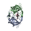


 Mass: 620.689 Da / Num. of mol.: 1 / Source method: obtained synthetically / Formula: C34H40N2O9
 Mass: 18.015 Da / Num. of mol.: 84 / Source method: isolated from a natural source / Formula: H2O
 Sample preparation
 / Beamline: I711 / Wavelength: 1.076
  Processing