 Movie
Movie Controller

[English] 日本語
 Yorodumi
- PDB-1wb8: Iron Superoxide Dismutase (FE-SOD) from the Hyperthermophile SULF... -
+ Open data

- Basic information
| Entry | Database: PDB / ID: 1wb8 | |||||||||
|---|---|---|---|---|---|---|---|---|---|---|
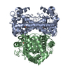
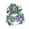
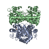
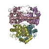
| Title | Iron Superoxide Dismutase (FE-SOD) from the Hyperthermophile SULFOLOBUS SOLFATARICUS. 2.3 A Resolution Structure of Recombinant Protein with a Covalently Modified Tyrosine in the Active Site. | |||||||||
 Components Components | SUPEROXIDE DISMUTASE [FE] | |||||||||
 Keywords Keywords |  OXIDOREDUCTASE / SUPEROXIDE DISMUTASE / SOD / IRON / SUPEROXIDE RADICAL DISPROPORTIONATION / SULFOLOBUS SOLFTARICUS / THERMOSTABLE OXIDOREDUCTASE / SUPEROXIDE DISMUTASE / SOD / IRON / SUPEROXIDE RADICAL DISPROPORTIONATION / SULFOLOBUS SOLFTARICUS / THERMOSTABLE | |||||||||
| Function / homology |  Function and homology informationsuperoxide dismutase / superoxide dismutase activity / metal ion binding / cytoplasm Function and homology informationsuperoxide dismutase / superoxide dismutase activity / metal ion binding / cytoplasmSimilarity search - Function | |||||||||
| Biological species |   SULFOLOBUS SOLFATARICUS (archaea) SULFOLOBUS SOLFATARICUS (archaea) | |||||||||
| Method | X-RAY DIFFRACTION / MOLECULAR REPLACEMENT / Resolution: 2.3 Å | |||||||||
 Authors Authors | Ursby, T. / Adinolfi, B.S. / Al-Karadaghi, S. / De Vendittis, E. / Bocchini, V. | |||||||||
 Citation Citation | Journal: J.Mol.Biol. / Year: 1999 Title: Iron Superoxide Dismutase from the Archaeon Sulfolobus Solfataricus: Analysis of Structure and Thermostability Authors: Ursby, T. / Adinolfi, B.S. / Al-Karadaghi, S. / De Vendittis, E. / Bocchini, V. #1: Journal: Eur.J.Biochem. / Year: 2001 Title: Phenylmethanesulfonyl Fluoride Inactivates an Archaeal Superoxide Dismutase by Chemical Modification of a Specific Tyrosine Residue Authors: De Vendittis, E. / Ursby, T. / Rullo, R. / Gogliettino, M.A. / Masullo, M. / Bocchini, V. | |||||||||
| History |
|
- Structure visualization
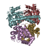
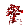
| Structure viewer | Molecule: MolmilJmol/JSmol |
|---|
- Downloads & links
-Download
| PDBx/mmCIF format | 1wb8.cif.gz | 99.1 KB | Display | PDBx/mmCIF format |
|---|---|---|---|---|
| PDB format | pdb1wb8.ent.gz | 75.5 KB | Display | PDB format |
| PDBx/mmJSON format | 1wb8.json.gz | Tree view | PDBx/mmJSON format | |
| Others |  Other downloads Other downloads |
-Validation report
| Arichive directory | https://data.pdbj.org/pub/pdb/validation_reports/wb/1wb8ftp://data.pdbj.org/pub/pdb/validation_reports/wb/1wb8 | HTTPS FTP |
|---|
-Related structure data
| Related structure data |  1abm S: Starting model for refinement |
|---|---|
| Similar structure data |


-Links
 PDBj
PDBj
- Assembly
| Deposited unit | 
| |||||||||
|---|---|---|---|---|---|---|---|---|---|---|
| 1 | 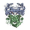
| |||||||||
| Unit cell |
| |||||||||
| Components on special symmetry positions |
| |||||||||
| Noncrystallographic symmetry (NCS) | NCS oper: (Code: given Matrix: (0.90666, -0.00174, 0.42185), Vector : |
-Components
| #1: Protein | Mass: 24142.312 Da / Num. of mol.: 2 Source method: isolated from a genetically manipulated source Details: COVALENT MODIFICATION OF TYR 41 CONSISTING OF HET GROUP PMS (PHENYL METHYL SULFONATE, BENZYLSULFINIC ACID) Source: (gene. exp.) SULFOLOBUS SOLFATARICUS (archaea) / Plasmid: PT7-7 / Production host:  ESCHERICHIA COLI (E. coli) / Strain (production host): JM109(DE3) / References: UniProt: P80857, superoxide dismutase ESCHERICHIA COLI (E. coli) / Strain (production host): JM109(DE3) / References: UniProt: P80857, superoxide dismutase#2: Chemical | Iron  Mass: 55.845 Da / Num. of mol.: 2 / Source method: obtained synthetically / Formula: Fe Mass: 55.845 Da / Num. of mol.: 2 / Source method: obtained synthetically / Formula: Fe#3: Chemical |   Mass: 172.202 Da / Num. of mol.: 2 / Source method: obtained synthetically / Formula: C7H8O3S Mass: 172.202 Da / Num. of mol.: 2 / Source method: obtained synthetically / Formula: C7H8O3S#4: Water | ChemComp-HOH / | Water Mass: 18.015 Da / Num. of mol.: 173 / Source method: isolated from a natural source / Formula: H2O Mass: 18.015 Da / Num. of mol.: 173 / Source method: isolated from a natural source / Formula: H2O |
|---|
-Experimental details
-Experiment
| Experiment | Method: X-RAY DIFFRACTION / Number of used crystals: 1 |
|---|
- Sample preparation
| Crystal | Density Matthews: 2.31 Å3/Da / Density % sol: 45 % |
|---|---|
| Crystal grow | Temperature: 294 K / Method: vapor diffusion, hanging drop / pH: 8.5 Details: CRYSTALS WERE GROWN BY VAPOR DIFFUSION IN HANGING DROPS AT 21C IN A 1 TO 1 MIX OF THE RESERVOIR SOLUTION (8% PEG 8000, 0.1 M TRIS/HCL PH 8.5) AND A PROTEIN SOLUTION (2.0 MG/ML SSSOD). |
-Data collection
| Diffraction | Mean temperature: 294 K |
|---|---|
| Diffraction source | Source: ROTATING ANODE / Type: RIGAKU ROTAFLEX RU-2 / Wavelength: 1.54 |
| Detector | Type: MARRESEARCH / Detector: IMAGE PLATE / Date: Jul 9, 1997 |
| Radiation | Monochromator: GRAPHITE / Protocol: SINGLE WAVELENGTH / Monochromatic (M) / Laue (L): M / Scattering type: x-ray |
| Radiation wavelength | Wavelength: 1.54 Å / Relative weight: 1 |
| Reflection | Resolution: 2.3→62 Å / Num. obs: 18647 / % possible obs: 96.3 % / Observed criterion σ(I): 0 / Redundancy: 4.2 % / Biso Wilson estimate: 14.9 Å2 / Rmerge(I) obs: 0.09 / Net I/σ(I): 7.4 |
| Reflection shell | Resolution: 2.3→2.36 Å / Redundancy: 4 % / Rmerge(I) obs: 0.31 / Mean I/σ(I) obs: 2.3 / % possible all: 94.7 |
- Processing
| Software |
| ||||||||||||||||||||||||||||||||||||||||||||||||||||||||||||||||||||||||||||||||
|---|---|---|---|---|---|---|---|---|---|---|---|---|---|---|---|---|---|---|---|---|---|---|---|---|---|---|---|---|---|---|---|---|---|---|---|---|---|---|---|---|---|---|---|---|---|---|---|---|---|---|---|---|---|---|---|---|---|---|---|---|---|---|---|---|---|---|---|---|---|---|---|---|---|---|---|---|---|---|---|---|---|
| Refinement | Method to determine structure: MOLECULAR REPLACEMENT Starting model: PDB ENTRY 1ABM 1abm Resolution: 2.3→22.91 Å / Rfactor Rfree error: 0.006 / Data cutoff high absF: 3370580.24 / Isotropic thermal model: RESTRAINED / Cross valid method: THROUGHOUT / σ(F): 0 Stereochemistry target values: MAXIMUM LIKELIHOOD USING AMPLITUDES Details: NOTE THAT RESIDUES 1-3 AND 209- -210 OF THE COMPLETE SEQUENCE ARE MISSING IN THIS ENTRY DUE TO DISORDER IN THE CRYSTAL. THE NCS USED IN THE REFINEMENT INCLUDED MOLECULE A EXCEPT 19 RESIDUES, ...Details: NOTE THAT RESIDUES 1-3 AND 209- -210 OF THE COMPLETE SEQUENCE ARE MISSING IN THIS ENTRY DUE TO DISORDER IN THE CRYSTAL. THE NCS USED IN THE REFINEMENT INCLUDED MOLECULE A EXCEPT 19 RESIDUES, THE IRON ION AND 68 WATER MOLECULES AND THE CORRESPONDING NCS RELEATED ATOMS. THE RMS DEVIATION FOR THE ATOMS USED FOR NCS IN THE REFINEMENT IS 0.006 A.
| ||||||||||||||||||||||||||||||||||||||||||||||||||||||||||||||||||||||||||||||||
| Solvent computation | Solvent model: FLAT MODEL / Bsol: 32.1856 Å2 / ksol: 0.294185 e/Å3 | ||||||||||||||||||||||||||||||||||||||||||||||||||||||||||||||||||||||||||||||||
| Displacement parameters | Biso mean: 23.3 Å2
| ||||||||||||||||||||||||||||||||||||||||||||||||||||||||||||||||||||||||||||||||
| Refine analyze |
| ||||||||||||||||||||||||||||||||||||||||||||||||||||||||||||||||||||||||||||||||
| Refinement step | Cycle: LAST / Resolution: 2.3→22.91 Å
| ||||||||||||||||||||||||||||||||||||||||||||||||||||||||||||||||||||||||||||||||
| Refine LS restraints |
| ||||||||||||||||||||||||||||||||||||||||||||||||||||||||||||||||||||||||||||||||
| LS refinement shell | Resolution: 2.3→2.44 Å / Rfactor Rfree error: 0.02 / Total num. of bins used: 6
| ||||||||||||||||||||||||||||||||||||||||||||||||||||||||||||||||||||||||||||||||
| Xplor file |
|
