 Movie
Movie Controller


+ Open data

- Basic information
| Entry | Database: PDB / ID: 1wac | ||||||
|---|---|---|---|---|---|---|---|
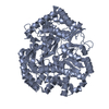
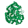
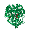
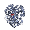
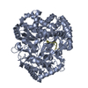
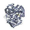
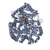
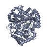
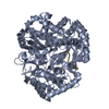
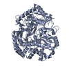
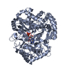
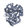
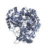
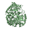
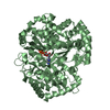
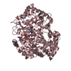
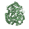
| Title | Back-priming mode of Phi6 RNA-dependent RNA polymerase | ||||||
 Components Components | P2 PROTEIN | ||||||
 Keywords Keywords |  POLYMERASE / PHI6 RNA-DEPENDENT RNA POLYMERASE / TRANSCRIPTION POLYMERASE / PHI6 RNA-DEPENDENT RNA POLYMERASE / TRANSCRIPTION | ||||||
| Function / homology |  Function and homology informationRNA uridylyltransferase activity / virion component / RNA-directed RNA polymerase / viral RNA genome replication / RNA-dependent RNA polymerase activity / nucleotide binding / DNA-templated transcription / RNA binding / metal ion binding Function and homology informationRNA uridylyltransferase activity / virion component / RNA-directed RNA polymerase / viral RNA genome replication / RNA-dependent RNA polymerase activity / nucleotide binding / DNA-templated transcription / RNA binding / metal ion bindingSimilarity search - Function | ||||||
| Biological species |  PSEUDOMONAS PHAGE PHI-6 (bacteriophage) PSEUDOMONAS PHAGE PHI-6 (bacteriophage) | ||||||
| Method | X-RAY DIFFRACTION / SYNCHROTRON / MOLECULAR REPLACEMENT / Resolution: 3 Å | ||||||
 Authors Authors | Laurila, M.R.L. / Salgado, P.S. / Stuart, D.I. / Grimes, J.M. / Bamford, D.H. | ||||||
 Citation Citation | Journal: J.Gen.Virol. / Year: 2005 Title: Back-Priming Mode of Phi6 RNA-Dependent RNA Polymerase Authors: Laurila, M.R.L. / Salgado, P.S. / Stuart, D.I. / Grimes, J.M. / Bamford, D.H. | ||||||
| History |
|
- Structure visualization
| Structure viewer | Molecule: MolmilJmol/JSmol |
|---|
- Downloads & links
-Download
| PDBx/mmCIF format | 1wac.cif.gz | 383.5 KB | Display | PDBx/mmCIF format |
|---|---|---|---|---|
| PDB format | pdb1wac.ent.gz | 316.4 KB | Display | PDB format |
| PDBx/mmJSON format | 1wac.json.gz | Tree view | PDBx/mmJSON format | |
| Others |  Other downloads Other downloads |
-Validation report
| Arichive directory | https://data.pdbj.org/pub/pdb/validation_reports/wa/1wacftp://data.pdbj.org/pub/pdb/validation_reports/wa/1wac | HTTPS FTP |
|---|
-Related structure data
| Related structure data | 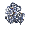 1hi8S S: Starting model for refinement |
|---|---|
| Similar structure data |


-Links
 PDBj
PDBj- Assembly
| Deposited unit | 
| ||||||||||||
|---|---|---|---|---|---|---|---|---|---|---|---|---|---|
| 1 | 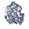
| ||||||||||||
| 2 | 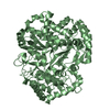
| ||||||||||||
| 3 | 
| ||||||||||||
| Unit cell |
| ||||||||||||
| Noncrystallographic symmetry (NCS) | NCS oper:
|
-Components
| #1: Protein | Mass: 74440.648 Da / Num. of mol.: 3 / Mutation: YES Source method: isolated from a genetically manipulated source Source: (gene. exp.) PSEUDOMONAS PHAGE PHI-6 (bacteriophage)Production host:  ESCHERICHIA COLI (E. coli) / References: UniProt: P11124 ESCHERICHIA COLI (E. coli) / References: UniProt: P11124Compound details | ENGINEERED RESIDUE MET 465 ILE, CHAINS A, B, C DELETION MUTATION- QYKW (629-632) TO SG IN CHAINS A, ...ENGINEERED | Sequence details | DELETION MUTATION- QYKW (629-632) TO SG IN CHAINS A, B AND C. THE TWO RESIDUES WHICH HAVE BEEN ...DELETION MUTATION- QYKW (629-632) TO SG IN CHAINS A, B AND C. THE TWO RESIDUES WHICH HAVE BEEN INSERTED IN PLACE OF QYKW ARE MAPPED TO THEMSELVES | |
|---|
-Experimental details
-Experiment
| Experiment | Method: X-RAY DIFFRACTION / Number of used crystals: 1 |
|---|
- Sample preparation
| Crystal | Density Matthews: 2.83 Å3/Da / Density % sol: 50 % |
|---|---|
| Crystal grow | pH: 8 Details: 0.1M SODIUM CITRATE PH 5.6, 19% ISOPROPANOL, 19% PEG 4K, 5% GLYCEROL |
-Data collection
| Diffraction | Mean temperature: 150 K |
|---|---|
| Diffraction source | Source: SYNCHROTRON / Site: ESRF  / Beamline: ID29 / Wavelength: 0.9756 / Beamline: ID29 / Wavelength: 0.9756 |
| Radiation | Protocol: SINGLE WAVELENGTH / Monochromatic (M) / Laue (L): M / Scattering type: x-ray |
| Radiation wavelength | Wavelength: 0.9756 Å / Relative weight: 1 |
| Reflection | Resolution: 3→30 Å / Num. obs: 57385 / % possible obs: 99.2 % / Observed criterion σ(I): 1.9 / Redundancy: 10.8 % / Biso Wilson estimate: 23.9 Å2 / Rmerge(I) obs: 0.02 / Net I/σ(I): 7.4 |
| Reflection shell | Resolution: 3→3.1 Å / Mean I/σ(I) obs: 1.9 / % possible all: 98.6 |
- Processing
| Software |
| ||||||||||||||||||||||||||||||||||||||||||||||||||||||||||||||||||||||||||||||||
|---|---|---|---|---|---|---|---|---|---|---|---|---|---|---|---|---|---|---|---|---|---|---|---|---|---|---|---|---|---|---|---|---|---|---|---|---|---|---|---|---|---|---|---|---|---|---|---|---|---|---|---|---|---|---|---|---|---|---|---|---|---|---|---|---|---|---|---|---|---|---|---|---|---|---|---|---|---|---|---|---|---|
| Refinement | Method to determine structure: MOLECULAR REPLACEMENT Starting model: PDB ENTRY 1HI8 Resolution: 3→20 Å / Rfactor Rfree error: 0.005 / Data cutoff high absF: 1769196.42 / Isotropic thermal model: RESTRAINED / Cross valid method: A POSTERIORI / σ(F): 0 Details: RESIDUES 1-5, 603-615, 626-634 BELONG TO DISORDERED LOOPS
| ||||||||||||||||||||||||||||||||||||||||||||||||||||||||||||||||||||||||||||||||
| Solvent computation | Solvent model: CNS BULK SOLVENT MODEL USED / Bsol: 45.2045 Å2 / ksol: 0.371779 e/Å3 | ||||||||||||||||||||||||||||||||||||||||||||||||||||||||||||||||||||||||||||||||
| Displacement parameters | Biso mean: 35.65 Å2
| ||||||||||||||||||||||||||||||||||||||||||||||||||||||||||||||||||||||||||||||||
| Refine analyze |
| ||||||||||||||||||||||||||||||||||||||||||||||||||||||||||||||||||||||||||||||||
| Refinement step | Cycle: LAST / Resolution: 3→20 Å
| ||||||||||||||||||||||||||||||||||||||||||||||||||||||||||||||||||||||||||||||||
| Refine LS restraints |
| ||||||||||||||||||||||||||||||||||||||||||||||||||||||||||||||||||||||||||||||||
| Refine LS restraints NCS | NCS model details: CONSTRAINTS | ||||||||||||||||||||||||||||||||||||||||||||||||||||||||||||||||||||||||||||||||
| LS refinement shell | Resolution: 3→3.19 Å / Rfactor Rfree error: 0.017 / Total num. of bins used: 6
|
