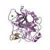
A: TRYPSIN INHIBITOR II B: TRYPSIN INHIBITOR II C: TRYPSIN INHIBITOR II D: TRYPSIN INHIBITOR II E: TRYPSIN INHIBITOR II F: TRYPSIN INHIBITOR II G: TRYPSIN INHIBITOR II H: TRYPSIN INHIBITOR II hetero molecules
A: TRYPSIN INHIBITOR II B: TRYPSIN INHIBITOR II C: TRYPSIN INHIBITOR II D: TRYPSIN INHIBITOR II E: TRYPSIN INHIBITOR II F: TRYPSIN INHIBITOR II hetero molecules
THE QUATERNARY STRUCTURE OF THE PROTEIN IS BELIEVED TOBE HEXAMERIC. CHAINS A TO F FORM THE HEXAMER, WHEREASCHAINS G AND H DO NOT OBEY ANY NON-CRYSTALLOGRAPHICSYMMETRY.
-
Components
#1: Protein/peptide
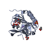
TRYPSININHIBITORII / / EETI-II
Mass: 3144.696 Da / Num. of mol.: 8 Source method: isolated from a genetically manipulated source Source: (gene. exp.) ECBALLIUM ELATERIUM (jumping cucumber) / Plasmid: PLZPWB-ETI-II / Production host: ESCHERICHIA COLI (E. coli) / Strain (production host): W3110 / References: UniProt: P12071
Resolution: 1.67→55.13 Å / Cor.coef. Fo:Fc: 0.95 / Cor.coef. Fo:Fc free: 0.929 / SU B: 4.394 / SU ML: 0.067 / Cross valid method: THROUGHOUT / ESU R: 0.112 / ESU R Free: 0.095 / Stereochemistry target values: MAXIMUM LIKELIHOOD / Details: HYDROGENS HAVE BEEN ADDED IN THE RIDING POSITIONS.
Rfactor
Num. reflection
% reflection
Selection details
Rfree
0.235
1779
5 %
RANDOM
Rwork
0.194
-
-
-
obs
0.197
33780
96.9 %
-
Solvent computation
Ion probe radii: 0.8 Å / Shrinkage radii: 0.8 Å / VDW probe radii: 1.2 Å / Solvent model: BABINET MODEL WITH MASK
 Movie
Movie Controller

 Yorodumi
 Open data
 Basic information
 Components
 Keywords
 Function and homology information
 Authors
 Citation
 Structure visualization
 Downloads & links
 Other downloads


 PDBj
PDBj
 Assembly


 Mass: 22.990 Da / Num. of mol.: 3 / Source method: obtained synthetically / Formula: Na
 Mass: 46.025 Da / Num. of mol.: 3 / Source method: obtained synthetically / Formula: CH2O2
 Mass: 18.015 Da / Num. of mol.: 130 / Source method: isolated from a natural source / Formula: H2O
 Sample preparation
 / Beamline: 14.2 / Wavelength: 0.9
  Processing