 Movie
Movie Controller

[English] 日本語
 Yorodumi
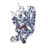
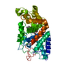
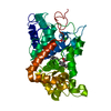
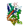
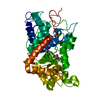
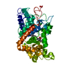
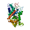
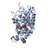
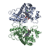
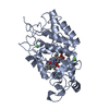
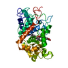
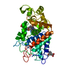
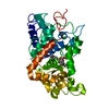
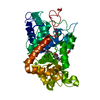
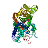
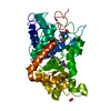
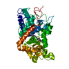
- PDB-1w4y: Ferrous horseradish peroxidase C1A in complex with carbon monoxide -
+ Open data

- Basic information
| Entry | Database: PDB / ID: 1w4y | ||||||
|---|---|---|---|---|---|---|---|
| Title | Ferrous horseradish peroxidase C1A in complex with carbon monoxide | ||||||
 Components Components | HORSERADISH PEROXIDASE C1A | ||||||
 Keywords Keywords |  OXIDOREDUCTASE / CARBON MONOXIDE / CALCIUM / FERROUS STATE / GLYCOPROTEIN / HEME / HORSERADISH / IRON / MULTIGENE FAMILY / PEROXIDASE / PYRROLIDONE CARBOXYLIC ACID OXIDOREDUCTASE / CARBON MONOXIDE / CALCIUM / FERROUS STATE / GLYCOPROTEIN / HEME / HORSERADISH / IRON / MULTIGENE FAMILY / PEROXIDASE / PYRROLIDONE CARBOXYLIC ACID | ||||||
| Function / homology |  Function and homology informationlactoperoxidase activity / peroxidase / vacuole / hydrogen peroxide catabolic process / response to oxidative stress / heme binding / extracellular region / metal ion binding Function and homology informationlactoperoxidase activity / peroxidase / vacuole / hydrogen peroxide catabolic process / response to oxidative stress / heme binding / extracellular region / metal ion bindingSimilarity search - Function | ||||||
| Biological species |  ARMORACIA RUSTICANA (horseradish) ARMORACIA RUSTICANA (horseradish) | ||||||
| Method | X-RAY DIFFRACTION / SYNCHROTRON / MOLECULAR REPLACEMENT / Resolution: 1.6 Å | ||||||
 Authors Authors | Carlsson, G.H. / Nicholls, P. / Svistunenko, D. / Berglund, G.I. / Hajdu, J. | ||||||
 Citation Citation | Journal: Biochemistry / Year: 2005 Title: Complexes of Horseradish Peroxidase with Formate, Acetate, and Carbon Monoxide Authors: Carlsson, G.H. / Nicholls, P. / Svistunenko, D. / Berglund, G.I. / Hajdu, J. #1: Journal: Nature / Year: 2002Title: The Catalytic Pathway of Horseradish Peroxidase at High Resolution Authors: Berglund, G.I. / Carlsson, G.H. / Smith, A.T. / Szoke, H. / Henriksen, A. / Hajdu, J. #2: Journal: J.Biol.Chem. / Year: 1990 Title: Expression of a Synthetic Gene for Horseradish Peroxidase C in Escherichia Coli and Folding and Activation of the Recombinant Enzyme with Ca2 and Heme Authors: Smith, A.T. / Santama, N. / Dacey, S. / Edwards, M. / Bray, R.C. / Thorneley, R.N. / Burke, J.F. | ||||||
| History |
|
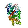
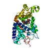
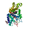
- Structure visualization
| Structure viewer | Molecule: MolmilJmol/JSmol |
|---|
- Downloads & links
-Download
| PDBx/mmCIF format | 1w4y.cif.gz | 84.6 KB | Display | PDBx/mmCIF format |
|---|---|---|---|---|
| PDB format | pdb1w4y.ent.gz | 61.1 KB | Display | PDB format |
| PDBx/mmJSON format | 1w4y.json.gz | Tree view | PDBx/mmJSON format | |
| Others |  Other downloads Other downloads |
-Validation report
| Arichive directory | https://data.pdbj.org/pub/pdb/validation_reports/w4/1w4yftp://data.pdbj.org/pub/pdb/validation_reports/w4/1w4y | HTTPS FTP |
|---|
-Related structure data
| Related structure data |  1w4wC  1h58S S: Starting model for refinement C: citing same article ( |
|---|---|
| Similar structure data |


-Links
 PDBj
PDBj


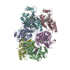
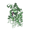
- Assembly
| Deposited unit | 
| ||||||||
|---|---|---|---|---|---|---|---|---|---|
| 1 |
| ||||||||
| Unit cell |
|
-Components
| #1: Protein | Mass: 35652.125 Da / Num. of mol.: 1 Source method: isolated from a genetically manipulated source Details: CARBON MONOXIDE BOUND TO THE FERROUS IRON / Source: (gene. exp.) ARMORACIA RUSTICANA (horseradish) / Production host:  ESCHERICHIA COLI (E. coli) / References: UniProt: P00433, peroxidase ESCHERICHIA COLI (E. coli) / References: UniProt: P00433, peroxidase | ||||
|---|---|---|---|---|---|
| #2: Chemical | ChemComp-HEM / Heme B  Mass: 616.487 Da / Num. of mol.: 1 / Source method: obtained synthetically / Formula: C34H32FeN4O4 Mass: 616.487 Da / Num. of mol.: 1 / Source method: obtained synthetically / Formula: C34H32FeN4O4 | ||||
| #3: Chemical |   Mass: 40.078 Da / Num. of mol.: 2 / Source method: obtained synthetically / Formula: Ca Mass: 40.078 Da / Num. of mol.: 2 / Source method: obtained synthetically / Formula: Ca#4: Chemical | ChemComp-CMO / | Carbon monoxide  Mass: 28.010 Da / Num. of mol.: 1 / Source method: obtained synthetically / Formula: CO Mass: 28.010 Da / Num. of mol.: 1 / Source method: obtained synthetically / Formula: CO#5: Water | ChemComp-HOH / | Water Mass: 18.015 Da / Num. of mol.: 341 / Source method: isolated from a natural source / Formula: H2O Mass: 18.015 Da / Num. of mol.: 341 / Source method: isolated from a natural source / Formula: H2O |
-Experimental details
-Experiment
| Experiment | Method: X-RAY DIFFRACTION / Number of used crystals: 1 |
|---|
- Sample preparation
| Crystal | Density Matthews: 2.24 Å3/Da / Density % sol: 45.2 % |
|---|---|
| Crystal grow | Temperature: 277 K / Method: vapor diffusion, hanging drop / pH: 6.5 Details: 20% PEG8000, 0.2 M CA-ACETATE, 0.1 M NA-CACODYLATE PH 6.5, FERULIC ACID AS ADDITIVE, HANGING DROP, STREAK SEEDING, 4DEGC |
-Data collection
| Diffraction | Mean temperature: 90 K |
|---|---|
| Diffraction source | Source: SYNCHROTRON / Site: MAX II  / Beamline: I711 / Wavelength: 1.089 / Beamline: I711 / Wavelength: 1.089 |
| Detector | Type: MARRESEARCH / Detector: CCD / Date: May 26, 2003 / Details: SI(III) |
| Radiation | Monochromator: MULTIPOLE WIGGLER / Protocol: SINGLE WAVELENGTH / Monochromatic (M) / Laue (L): M / Scattering type: x-ray |
| Radiation wavelength | Wavelength: 1.089 Å / Relative weight: 1 |
| Reflection | Resolution: 1.57→50 Å / Num. obs: 41633 / % possible obs: 90.4 % / Observed criterion σ(I): 3 / Redundancy: 2.9 % / Biso Wilson estimate: 17.9 Å2 / Rmerge(I) obs: 0.07 / Net I/σ(I): 8.3 |
| Reflection shell | Resolution: 1.57→1.61 Å / Rmerge(I) obs: 0.3 / % possible all: 86.8 |
- Processing
| Software |
| ||||||||||||||||||||||||||||||||||||||||||||||||||||||||||||||||||||||||||||||||
|---|---|---|---|---|---|---|---|---|---|---|---|---|---|---|---|---|---|---|---|---|---|---|---|---|---|---|---|---|---|---|---|---|---|---|---|---|---|---|---|---|---|---|---|---|---|---|---|---|---|---|---|---|---|---|---|---|---|---|---|---|---|---|---|---|---|---|---|---|---|---|---|---|---|---|---|---|---|---|---|---|---|
| Refinement | Method to determine structure: MOLECULAR REPLACEMENT Starting model: PDB ENTRY 1H58 Resolution: 1.6→25.94 Å / Rfactor Rfree error: 0.005 / Data cutoff high absF: 1352591.14 / Isotropic thermal model: RESTRAINED / Cross valid method: THROUGHOUT / σ(F): 0 / Stereochemistry target values: MAXIMUM LIKELIHOOD
| ||||||||||||||||||||||||||||||||||||||||||||||||||||||||||||||||||||||||||||||||
| Solvent computation | Solvent model: FLAT MODEL / Bsol: 44.2791 Å2 / ksol: 0.371891 e/Å3 | ||||||||||||||||||||||||||||||||||||||||||||||||||||||||||||||||||||||||||||||||
| Displacement parameters | Biso mean: 16.4 Å2
| ||||||||||||||||||||||||||||||||||||||||||||||||||||||||||||||||||||||||||||||||
| Refine analyze |
| ||||||||||||||||||||||||||||||||||||||||||||||||||||||||||||||||||||||||||||||||
| Refinement step | Cycle: LAST / Resolution: 1.6→25.94 Å
| ||||||||||||||||||||||||||||||||||||||||||||||||||||||||||||||||||||||||||||||||
| Refine LS restraints |
| ||||||||||||||||||||||||||||||||||||||||||||||||||||||||||||||||||||||||||||||||
| LS refinement shell | Resolution: 1.6→1.7 Å / Rfactor Rfree error: 0.015 / Total num. of bins used: 6
| ||||||||||||||||||||||||||||||||||||||||||||||||||||||||||||||||||||||||||||||||
| Xplor file |
|
