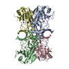
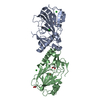
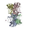
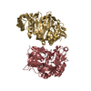
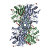
[English] 日本語
Yorodumi
- PDB-1vz4: Fe-Succinate Complex of AtsK -

+
Open data


ID or keywords:

Loading...

-
Basic information

Entry
Database: PDB / ID: 1vz4
TitleFe-Succinate Complex of AtsK
ComponentsPUTATIVE ALKYLSULFATASE ATSK
KeywordsOXIDOREDUCTASE / NON-HEME FE(II) ALPHAKETOGLUTARATE DEPENDENT DIOXYGENASE / ALKYLSULFATASE / JELLY ROLL / OXIDOREDUCTASE SULFATASE / SELF HYDROXYLATION
Function / homology
Function and homology information


alkyl sulfatase / dioxygenase activity / metal ion binding
Similarity search - Function
Clavaminate synthase-like / Double-stranded beta-helix / TauD/TfdA-like domain / Taurine catabolism dioxygenase TauD, TfdA family / Taurine dioxygenase TauD-like superfamily / 4-Layer Sandwich / Alpha Beta
Similarity search - Domain/homology
: / SUCCINIC ACID / Alpha-ketoglutarate-dependent sulfate ester dioxygenase
Similarity search - Component
Biological speciesPSEUDOMONAS PUTIDA (bacteria)
MethodX-RAY DIFFRACTION / SYNCHROTRON / MOLECULAR REPLACEMENT / Resolution: 2.5 Å
AuthorsMueller, I. / Stueckl, A.C. / Uson, I. / Kertesz, M.
CitationJournal: J.Biol.Chem. / Year: 2005
Title: Succinate Complex Crystal Structures of the Alpha-Ketoglutarate-Dependent Dioxygenase Atsk: Steric Aspects of Enzyme Self-Hydroxylation
Authors: Mueller, I. / Stueckl, A.C. / Wakeley, J. / Kertesz, M. / Uson, I.
History
DepositionMay 14, 2004Deposition site: PDBE / Processing site: PDBE
Revision 1.0Nov 15, 2004Provider: repository / Type: Initial release
Revision 1.1May 8, 2011Group: Version format compliance
Revision 1.2Jul 13, 2011Group: Version format compliance
Revision 1.3Dec 13, 2023Group: Data collection / Database references ...Data collection / Database references / Derived calculations / Other / Refinement description
Category: chem_comp_atom / chem_comp_bond ...chem_comp_atom / chem_comp_bond / database_2 / pdbx_database_status / pdbx_initial_refinement_model / pdbx_struct_conn_angle / struct_conn
Item: _database_2.pdbx_DOI / _database_2.pdbx_database_accession ..._database_2.pdbx_DOI / _database_2.pdbx_database_accession / _pdbx_database_status.status_code_sf / _pdbx_struct_conn_angle.ptnr1_auth_comp_id / _pdbx_struct_conn_angle.ptnr1_auth_seq_id / _pdbx_struct_conn_angle.ptnr1_label_asym_id / _pdbx_struct_conn_angle.ptnr1_label_atom_id / _pdbx_struct_conn_angle.ptnr1_label_comp_id / _pdbx_struct_conn_angle.ptnr1_label_seq_id / _pdbx_struct_conn_angle.ptnr3_auth_comp_id / _pdbx_struct_conn_angle.ptnr3_auth_seq_id / _pdbx_struct_conn_angle.ptnr3_label_asym_id / _pdbx_struct_conn_angle.ptnr3_label_atom_id / _pdbx_struct_conn_angle.ptnr3_label_comp_id / _pdbx_struct_conn_angle.ptnr3_label_seq_id / _pdbx_struct_conn_angle.value / _struct_conn.pdbx_dist_value / _struct_conn.ptnr1_auth_comp_id / _struct_conn.ptnr1_auth_seq_id / _struct_conn.ptnr1_label_asym_id / _struct_conn.ptnr1_label_atom_id / _struct_conn.ptnr1_label_comp_id / _struct_conn.ptnr1_label_seq_id / _struct_conn.ptnr2_auth_comp_id / _struct_conn.ptnr2_auth_seq_id / _struct_conn.ptnr2_label_asym_id / _struct_conn.ptnr2_label_atom_id / _struct_conn.ptnr2_label_comp_id / _struct_conn.ptnr2_label_seq_id
Remark 700 SHEET THE SHEET STRUCTURE OF THIS MOLECULE IS BIFURCATED. IN ORDER TO REPRESENT THIS FEATURE IN ... SHEET THE SHEET STRUCTURE OF THIS MOLECULE IS BIFURCATED. IN ORDER TO REPRESENT THIS FEATURE IN THE SHEET RECORDS BELOW, TWO SHEETS ARE DEFINED.

-
Structure visualization

Structure viewerMolecule:
MolmilJmol/JSmol

Downloads & links

-
Assembly

Deposited unit
A: PUTATIVE ALKYLSULFATASE ATSK
D: PUTATIVE ALKYLSULFATASE ATSK
hetero molecules


Theoretical massNumber of molelcules
Total (without water)66,8456
Polymers66,4972
Non-polymers3484
Water86548
1
A: PUTATIVE ALKYLSULFATASE ATSK
D: PUTATIVE ALKYLSULFATASE ATSK
hetero molecules

A: PUTATIVE ALKYLSULFATASE ATSK
D: PUTATIVE ALKYLSULFATASE ATSK
hetero molecules


Theoretical massNumber of molelcules
Total (without water)133,68912
Polymers132,9944
Non-polymers6968
Water724
TypeNameSymmetry operationNumber
identity operation1_555x,y,z1
crystal symmetry operation8_565x,-y+1,-z+1/21
MethodPQS
Unit cell
Length a, b, c (Å)71.845, 141.000, 160.149
Angle α, β, γ (deg.)90.00, 90.00, 90.00
Int Tables number24
Space group name H-MI212121
Noncrystallographic symmetry (NCS)NCS domain:
IDEns-IDDetails
11A
21D

NCS domain segments:
Dom-IDComponent-IDEns-IDRefine codeAuth asym-IDAuth seq-ID
1112A13 - 80
2112D13 - 80
1212A112 - 164
2212D112 - 164
1312A191 - 213
2312D191 - 213
1412A215 - 263
2412D215 - 263
1512A280 - 298
2512D280 - 298

-
Components

#1: Protein PUTATIVE ALKYLSULFATASE ATSK


Mass: 33248.402 Da / Num. of mol.: 2 / Source method: isolated from a natural source / Source: (natural) PSEUDOMONAS PUTIDA (bacteria) / Strain: S-313 / References: UniProt: Q9WWU5
#2: Chemical ChemComp-FE2 / FE (II) ION


Mass: 55.845 Da / Num. of mol.: 2 / Source method: obtained synthetically / Formula: Fe
#3: Chemical ChemComp-SIN / SUCCINIC ACID / Succinic acid


Mass: 118.088 Da / Num. of mol.: 2 / Source method: obtained synthetically / Formula: C4H6O4
#4: Water ChemComp-HOH / water / Water


Mass: 18.015 Da / Num. of mol.: 48 / Source method: isolated from a natural source / Formula: H2O

-
Experimental details

-
Experiment

ExperimentMethod: X-RAY DIFFRACTION / Number of used crystals: 1

-
Sample preparation

CrystalDensity Matthews: 3.05 Å3/Da / Density % sol: 60 %
Description: SIDE CHAINS IN ACTIVE SITE TRUNCATED FOR MOLECULAR REPLACEMENT MODEL
Crystal growpH: 7
Details: 25 MM AMMONIUM SULFATE 175 MM SODUIM SUCCINATE PH 5.6, 15 % PEG 4000 30 % GLYCEROL SOAKING IN 2 MM FE(NH4)2(SO4)2X6H2O

-
Data collection

DiffractionMean temperature: 100 K
Diffraction sourceSource: SYNCHROTRON / Site: BESSY / Beamline: 14.2 / Wavelength: 0.9
DetectorType: MARRESEARCH / Detector: IMAGE PLATE / Details: MIRRORS
RadiationProtocol: SINGLE WAVELENGTH / Monochromatic (M) / Laue (L): M / Scattering type: x-ray
Radiation wavelengthWavelength: 0.9 Å / Relative weight: 1
ReflectionResolution: 2.5→50 Å / Num. obs: 27572 / % possible obs: 95.9 % / Redundancy: 3.8 % / Rmerge(I) obs: 0.081 / Net I/σ(I): 24
Reflection shellResolution: 2.5→2.6 Å / Redundancy: 2.1 % / Rmerge(I) obs: 0.128 / Mean I/σ(I) obs: 9.53 / % possible all: 76.4

-
Processing

Software
NameVersionClassification
REFMAC5.2.0003refinement
DENZOdata reduction
SCALEPACKdata scaling
RefinementMethod to determine structure: MOLECULAR REPLACEMENT
Starting model: PDB ENTRY 1OIH
Resolution: 2.5→50 Å / Cor.coef. Fo:Fc: 0.935 / Cor.coef. Fo:Fc free: 0.907 / SU B: 10.36 / SU ML: 0.229 / Cross valid method: THROUGHOUT / ESU R: 0.362 / ESU R Free: 0.286 / Stereochemistry target values: MAXIMUM LIKELIHOOD
RfactorNum. reflection% reflectionSelection details
Rfree0.287 1461 5.4 %RANDOM
Rwork0.227 ---
obs0.23 25828 99.2 %-
Solvent computationIon probe radii: 0.8 Å / Shrinkage radii: 0.8 Å / VDW probe radii: 1.2 Å / Solvent model: BABINET MODEL WITH MASK
Displacement parametersBiso mean: 50.06 Å2
Baniso -1Baniso -2Baniso -3
1--6.09 Å20 Å20 Å2
2---4.26 Å20 Å2
3---10.35 Å2
Refinement stepCycle: LAST / Resolution: 2.5→50 Å
ProteinNucleic acidLigandSolventTotal
Num. atoms3871 0 18 48 3937
Refine LS restraints
Refine-IDTypeDev idealDev ideal targetNumber
X-RAY DIFFRACTIONr_bond_refined_d0.0290.0213975
X-RAY DIFFRACTIONr_bond_other_d
X-RAY DIFFRACTIONr_angle_refined_deg2.4181.9165430
X-RAY DIFFRACTIONr_angle_other_deg
X-RAY DIFFRACTIONr_dihedral_angle_1_deg8.3835500
X-RAY DIFFRACTIONr_dihedral_angle_2_deg29.25522.912182
X-RAY DIFFRACTIONr_dihedral_angle_3_deg18.80215560
X-RAY DIFFRACTIONr_dihedral_angle_4_deg17.3111533
X-RAY DIFFRACTIONr_chiral_restr0.1610.2620
X-RAY DIFFRACTIONr_gen_planes_refined0.0110.023081
X-RAY DIFFRACTIONr_gen_planes_other
X-RAY DIFFRACTIONr_nbd_refined0.2420.21727
X-RAY DIFFRACTIONr_nbd_other
X-RAY DIFFRACTIONr_nbtor_refined0.3170.22611
X-RAY DIFFRACTIONr_nbtor_other
X-RAY DIFFRACTIONr_xyhbond_nbd_refined0.1720.2156
X-RAY DIFFRACTIONr_xyhbond_nbd_other
X-RAY DIFFRACTIONr_metal_ion_refined0.0130.21
X-RAY DIFFRACTIONr_metal_ion_other
X-RAY DIFFRACTIONr_symmetry_vdw_refined0.2630.227
X-RAY DIFFRACTIONr_symmetry_vdw_other
X-RAY DIFFRACTIONr_symmetry_hbond_refined0.2380.27
X-RAY DIFFRACTIONr_symmetry_hbond_other
X-RAY DIFFRACTIONr_symmetry_metal_ion_refined
X-RAY DIFFRACTIONr_symmetry_metal_ion_other
X-RAY DIFFRACTIONr_mcbond_it2.11722611
X-RAY DIFFRACTIONr_mcbond_other
X-RAY DIFFRACTIONr_mcangle_it3.47444014
X-RAY DIFFRACTIONr_mcangle_other
X-RAY DIFFRACTIONr_scbond_it4.58741608
X-RAY DIFFRACTIONr_scbond_other
X-RAY DIFFRACTIONr_scangle_it6.08761416
X-RAY DIFFRACTIONr_scangle_other
X-RAY DIFFRACTIONr_long_range_B_refined
X-RAY DIFFRACTIONr_long_range_B_other
X-RAY DIFFRACTIONr_rigid_bond_restr
X-RAY DIFFRACTIONr_sphericity_free
X-RAY DIFFRACTIONr_sphericity_bonded
Refine LS restraints NCS

Dom-ID: 1 / Auth asym-ID: A / Ens-ID: 1 / Refine-ID: X-RAY DIFFRACTION

NumberTypeRms dev position (Å)Weight position
844tight positional0.10.05
719medium positional0.310.5
844tight thermal0.430.5
719medium thermal2.242
LS refinement shellResolution: 2.5→2.56 Å / Total num. of bins used: 20
RfactorNum. reflection% reflection
Rfree0.438 191 -
Rwork0.301 1397 -
obs--99.81 %

+
About Yorodumi

-
News

-
Feb 9, 2022. New format data for meta-information of EMDB entries

New format data for meta-information of EMDB entries

  • Version 3 of the EMDB header file is now the official format.
  • The previous official version 1.9 will be removed from the archive.

Related info.:EMDB header

External links:wwPDB to switch to version 3 of the EMDB data model

-
Aug 12, 2020. Covid-19 info

Covid-19 info

URL: https://pdbj.org/emnavi/covid19.php

New page: Covid-19 featured information page in EM Navigator.

Related info.:Covid-19 info / Mar 5, 2020. Novel coronavirus structure data

+
Mar 5, 2020. Novel coronavirus structure data

Novel coronavirus structure data

Related info.:Yorodumi Speices / Aug 12, 2020. Covid-19 info

External links:COVID-19 featured content - PDBj / Molecule of the Month (242):Coronavirus Proteases

+
Jan 31, 2019. EMDB accession codes are about to change! (news from PDBe EMDB page)

EMDB accession codes are about to change! (news from PDBe EMDB page)

  • The allocation of 4 digits for EMDB accession codes will soon come to an end. Whilst these codes will remain in use, new EMDB accession codes will include an additional digit and will expand incrementally as the available range of codes is exhausted. The current 4-digit format prefixed with “EMD-” (i.e. EMD-XXXX) will advance to a 5-digit format (i.e. EMD-XXXXX), and so on. It is currently estimated that the 4-digit codes will be depleted around Spring 2019, at which point the 5-digit format will come into force.
  • The EM Navigator/Yorodumi systems omit the EMD- prefix.

Related info.:Q: What is EMD? / ID/Accession-code notation in Yorodumi/EM Navigator

External links:EMDB Accession Codes are Changing Soon! / Contact to PDBj

+
Jul 12, 2017. Major update of PDB

Major update of PDB

  • wwPDB released updated PDB data conforming to the new PDBx/mmCIF dictionary.
  • This is a major update changing the version number from 4 to 5, and with Remediation, in which all the entries are updated.
  • In this update, many items about electron microscopy experimental information are reorganized (e.g. em_software).
  • Now, EM Navigator and Yorodumi are based on the updated data.

External links:wwPDB Remediation / Enriched Model Files Conforming to OneDep Data Standards Now Available in the PDB FTP Archive

-
Yorodumi

Thousand views of thousand structures

  • Yorodumi is a browser for structure data from EMDB, PDB, SASBDB, etc.
  • This page is also the successor to EM Navigator detail page, and also detail information page/front-end page for Omokage search.
  • The word "yorodu" (or yorozu) is an old Japanese word meaning "ten thousand". "mi" (miru) is to see.

Related info.:EMDB / PDB / SASBDB / Comparison of 3 databanks / Yorodumi Search / Aug 31, 2016. New EM Navigator & Yorodumi / Yorodumi Papers / Jmol/JSmol / Function and homology information / Changes in new EM Navigator and Yorodumi

Read more