 Movie
Movie Controller

[English] 日本語
 Yorodumi
- PDB-1vr9: CRYSTAL STRUCTURE OF A CBS DOMAIN PAIR/ACT DOMAIN PROTEIN (TM0892... -
+ Open data

- Basic information
| Entry | Database: PDB / ID: 1vr9 | ||||||
|---|---|---|---|---|---|---|---|
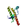
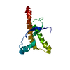
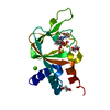
| Title | CRYSTAL STRUCTURE OF A CBS DOMAIN PAIR/ACT DOMAIN PROTEIN (TM0892) FROM THERMOTOGA MARITIMA AT 1.70 A RESOLUTION | ||||||
 Components Components | CBS domain protein/ACT domain protein | ||||||
 Keywords Keywords | UNKNOWN FUNCTION / STRUCTURAL GENOMICS / JOINT CENTER FOR STRUCTURAL GENOMICS / JCSG / PROTEIN STRUCTURE INITIATIVE / PSI | ||||||
| Function / homology |  Function and homology information Function and homology information | ||||||
| Biological species |   Thermotoga maritima (bacteria) Thermotoga maritima (bacteria) | ||||||
| Method | X-RAY DIFFRACTION / SYNCHROTRON / MAD, MOLECULAR REPLACEMENT / Resolution: 1.7 Å | ||||||
 Authors Authors | Joint Center for Structural Genomics (JCSG) | ||||||
 Citation Citation | Journal: To be published Title: Crystal structure of CBS domain protein/ACT domain protein (TM0892) from Thermotoga maritima at 1.70 A resolution Authors: Joint Center for Structural Genomics (JCSG) | ||||||
| History |
|
- Structure visualization
| Structure viewer | Molecule: MolmilJmol/JSmol |
|---|
- Downloads & links
-Download
| PDBx/mmCIF format | 1vr9.cif.gz | 67.7 KB | Display | PDBx/mmCIF format |
|---|---|---|---|---|
| PDB format | pdb1vr9.ent.gz | 52.4 KB | Display | PDB format |
| PDBx/mmJSON format | 1vr9.json.gz | Tree view | PDBx/mmJSON format | |
| Others |  Other downloads Other downloads |
-Validation report
| Arichive directory | https://data.pdbj.org/pub/pdb/validation_reports/vr/1vr9ftp://data.pdbj.org/pub/pdb/validation_reports/vr/1vr9 | HTTPS FTP |
|---|
-Related structure data
| Similar structure data | |
|---|---|
| Other databases |


-Links
 PDBj
PDBj


- Assembly
| Deposited unit | 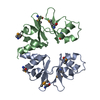
| ||||||||
|---|---|---|---|---|---|---|---|---|---|
| 1 | 
| ||||||||
| 2 | 
| ||||||||
| Unit cell |
|
-Components
| #1: Protein | Mass: 24665.336 Da / Num. of mol.: 2 Source method: isolated from a genetically manipulated source Source: (gene. exp.) Thermotoga maritima (bacteria) / Gene: TM0892 / Production host: Escherichia coli (E. coli) / References: UniProt: Q9WZZ4#2: Water | ChemComp-HOH / | Water Mass: 18.015 Da / Num. of mol.: 242 / Source method: isolated from a natural source / Formula: H2O Mass: 18.015 Da / Num. of mol.: 242 / Source method: isolated from a natural source / Formula: H2O |
|---|
-Experimental details
-Experiment
| Experiment | Method: X-RAY DIFFRACTION / Number of used crystals: 2 |
|---|
- Sample preparation
| Crystal |
| |||||||||||||||
|---|---|---|---|---|---|---|---|---|---|---|---|---|---|---|---|---|
| Crystal grow |
|
-Data collection
| Diffraction |
| ||||||||||||||||||
|---|---|---|---|---|---|---|---|---|---|---|---|---|---|---|---|---|---|---|---|
| Diffraction source |
| ||||||||||||||||||
| Detector |
| ||||||||||||||||||
| Radiation |
| ||||||||||||||||||
| Radiation wavelength |
| ||||||||||||||||||
| Reflection | Resolution: 1.7→37.81 Å / Num. obs: 45259 / % possible obs: 81.56 % / Redundancy: 3.16 % / Biso Wilson estimate: 29.22 Å2 / Rmerge(I) obs: 0.088 / Net I/σ(I): 20.66 | ||||||||||||||||||
| Reflection shell | Resolution: 1.7→1.76 Å / Redundancy: 1.96 % / Rmerge(I) obs: 0.278 / Mean I/σ(I) obs: 2.45 / Num. unique all: 4044 / % possible all: 73.13 |

- Processing
| Software |
| |||||||||||||||||||||||||||||||||||||||||||||||||||||||||||||||||||||||||||||||||||||||||||||||||||||||||||||||||||||||||||||
|---|---|---|---|---|---|---|---|---|---|---|---|---|---|---|---|---|---|---|---|---|---|---|---|---|---|---|---|---|---|---|---|---|---|---|---|---|---|---|---|---|---|---|---|---|---|---|---|---|---|---|---|---|---|---|---|---|---|---|---|---|---|---|---|---|---|---|---|---|---|---|---|---|---|---|---|---|---|---|---|---|---|---|---|---|---|---|---|---|---|---|---|---|---|---|---|---|---|---|---|---|---|---|---|---|---|---|---|---|---|---|---|---|---|---|---|---|---|---|---|---|---|---|---|---|---|---|
| Refinement | Method to determine structure: MAD, MOLECULAR REPLACEMENT / Resolution: 1.7→37.81 Å / Cor.coef. Fo:Fc: 0.906 / Cor.coef. Fo:Fc free: 0.895 / SU B: 3.434 / SU ML: 0.06 / TLS residual ADP flag: LIKELY RESIDUAL / Cross valid method: THROUGHOUT / ESU R: 0.117 / ESU R Free: 0.115 / Stereochemistry target values: MAXIMUM LIKELIHOOD Details: 1.HYDROGENS HAVE BEEN ADDED IN THE RIDING POSITIONS. 2. IT WAS NOT POSSIBLE TO TRACE RESIDUES 122-201 IN EACH CHAIN. PFAM ANALYSIS OF THE SEQUENCE INDICATES THAT THIS REGION IS COMPOSED OF ...Details: 1.HYDROGENS HAVE BEEN ADDED IN THE RIDING POSITIONS. 2. IT WAS NOT POSSIBLE TO TRACE RESIDUES 122-201 IN EACH CHAIN. PFAM ANALYSIS OF THE SEQUENCE INDICATES THAT THIS REGION IS COMPOSED OF AN ACT DOMAIN. MASS SPECTROSCOPY INDICATES THAT THE PROTEIN IS COMPLETE, SUGGESTING THAT THIS REGION IS DISORDERED.
| |||||||||||||||||||||||||||||||||||||||||||||||||||||||||||||||||||||||||||||||||||||||||||||||||||||||||||||||||||||||||||||
| Solvent computation | Ion probe radii: 0.8 Å / Shrinkage radii: 0.8 Å / VDW probe radii: 1.2 Å / Solvent model: MASK | |||||||||||||||||||||||||||||||||||||||||||||||||||||||||||||||||||||||||||||||||||||||||||||||||||||||||||||||||||||||||||||
| Displacement parameters | Biso mean: 23.729 Å2
| |||||||||||||||||||||||||||||||||||||||||||||||||||||||||||||||||||||||||||||||||||||||||||||||||||||||||||||||||||||||||||||
| Refinement step | Cycle: LAST / Resolution: 1.7→37.81 Å
| |||||||||||||||||||||||||||||||||||||||||||||||||||||||||||||||||||||||||||||||||||||||||||||||||||||||||||||||||||||||||||||
| Refine LS restraints |
| |||||||||||||||||||||||||||||||||||||||||||||||||||||||||||||||||||||||||||||||||||||||||||||||||||||||||||||||||||||||||||||
| LS refinement shell | Resolution: 1.697→1.741 Å / Total num. of bins used: 20
| |||||||||||||||||||||||||||||||||||||||||||||||||||||||||||||||||||||||||||||||||||||||||||||||||||||||||||||||||||||||||||||
| Refinement TLS params. | Method: refined / Refine-ID: X-RAY DIFFRACTION
| |||||||||||||||||||||||||||||||||||||||||||||||||||||||||||||||||||||||||||||||||||||||||||||||||||||||||||||||||||||||||||||
| Refinement TLS group | Refine-ID: X-RAY DIFFRACTION / Selection: ALL / Auth seq-ID: 1 - 121 / Label seq-ID: 13 - 133
|
