 Movie
Movie Controller

[English] 日本語
 Yorodumi
- PDB-1vq3: Crystal structure of Phosphoribosylformylglycinamidine synthase, ... -
+ Open data

- Basic information
| Entry | Database: PDB / ID: 1vq3 | ||||||
|---|---|---|---|---|---|---|---|
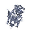
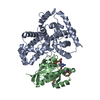
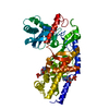
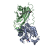
| Title | Crystal structure of Phosphoribosylformylglycinamidine synthase, purS subunit (EC 6.3.5.3) (TM1244) from Thermotoga maritima at 1.90 A resolution | ||||||
 Components Components | Phosphoribosylformylglycinamidine synthase, purS subunit | ||||||
 Keywords Keywords | LIGASE / TM1244 / phosphoribosylformylglycinamidine synthase / purs subunit (EC 6.3.5.3) / Structural Genomics / Joint Center for Structural Genomics / JCSG / Protein Structure Initiative / PSI | ||||||
| Function / homology |  Function and homology informationphosphoribosylformylglycinamidine synthase / phosphoribosylformylglycinamidine synthase activity / 'de novo' IMP biosynthetic process / ATP binding / cytoplasm Function and homology informationphosphoribosylformylglycinamidine synthase / phosphoribosylformylglycinamidine synthase activity / 'de novo' IMP biosynthetic process / ATP binding / cytoplasmSimilarity search - Function | ||||||
| Biological species |   Thermotoga maritima (bacteria) Thermotoga maritima (bacteria) | ||||||
| Method | X-RAY DIFFRACTION / SYNCHROTRON / MOLECULAR REPLACEMENT / Resolution: 1.9 Å | ||||||
 Authors Authors | Joint Center for Structural Genomics (JCSG) | ||||||
 Citation Citation | Journal: Proteins / Year: 2006 Title: Crystal structure of phosphoribosylformyl-glycinamidine synthase II, PurS subunit (TM1244) from Thermotoga maritima at 1.90 A resolution. Authors: Mathews, I.I. / Krishna, S.S. / Schwarzenbacher, R. / McMullan, D. / Jaroszewski, L. / Miller, M.D. / Abdubek, P. / Agarwalla, S. / Ambing, E. / Axelrod, H.L. / Canaves, J.M. / Carlton, D. / ...Authors: Mathews, I.I. / Krishna, S.S. / Schwarzenbacher, R. / McMullan, D. / Jaroszewski, L. / Miller, M.D. / Abdubek, P. / Agarwalla, S. / Ambing, E. / Axelrod, H.L. / Canaves, J.M. / Carlton, D. / Chiu, H.J. / Clayton, T. / DiDonato, M. / Duan, L. / Elsliger, M.A. / Grzechnik, S.K. / Hale, J. / Hampton, E. / Haugen, J. / Jin, K.K. / Klock, H.E. / Koesema, E. / Kovarik, J.S. / Kreusch, A. / Kuhn, P. / Levin, I. / Morse, A.T. / Nigoghossian, E. / Okach, L. / Oommachen, S. / Paulsen, J. / Quijano, K. / Reyes, R. / Rife, C.L. / Spraggon, G. / Stevens, R.C. / van den Bedem, H. / White, A. / Wolf, G. / Xu, Q. / Hodgson, K.O. / Wooley, J. / Deacon, A.M. / Godzik, A. / Lesley, S.A. / Wilson, I.A. | ||||||
| History |
| ||||||
| Remark 999 | SEQUENCE CLONING ARTIFACT: THIS GENE USES AN ALTERNATE INITIATION CODON THAT RESULTS IN A LEUCINE ... SEQUENCE CLONING ARTIFACT: THIS GENE USES AN ALTERNATE INITIATION CODON THAT RESULTS IN A LEUCINE AT POSITION 1 WHEN EXPRESSED AS A FUSION WITH THE PURIFICATION TAG |
- Structure visualization
| Structure viewer | Molecule: MolmilJmol/JSmol |
|---|
- Downloads & links
-Download
| PDBx/mmCIF format | 1vq3.cif.gz | 87.7 KB | Display | PDBx/mmCIF format |
|---|---|---|---|---|
| PDB format | pdb1vq3.ent.gz | 66.7 KB | Display | PDB format |
| PDBx/mmJSON format | 1vq3.json.gz | Tree view | PDBx/mmJSON format | |
| Others |  Other downloads Other downloads |
-Validation report
| Arichive directory | https://data.pdbj.org/pub/pdb/validation_reports/vq/1vq3ftp://data.pdbj.org/pub/pdb/validation_reports/vq/1vq3 | HTTPS FTP |
|---|
-Related structure data
| Related structure data |  1t4aS S: Starting model for refinement |
|---|---|
| Similar structure data | |
| Other databases |


-Links
 PDBj
PDBj- Assembly
| Deposited unit | 
| ||||||||||||||||||||||||||||||
|---|---|---|---|---|---|---|---|---|---|---|---|---|---|---|---|---|---|---|---|---|---|---|---|---|---|---|---|---|---|---|---|
| 1 |
| ||||||||||||||||||||||||||||||
| 2 | 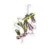
| ||||||||||||||||||||||||||||||
| 3 | 
| ||||||||||||||||||||||||||||||
| Unit cell |
| ||||||||||||||||||||||||||||||
| Noncrystallographic symmetry (NCS) | NCS domain:
NCS domain segments: Component-ID: 1 / Ens-ID: 1 / Beg auth comp-ID: PHE / Beg label comp-ID: PHE / End auth comp-ID: LEU / End label comp-ID: LEU / Refine code: 4 / Auth seq-ID: 4 - 82 / Label seq-ID: 16 - 94
|
-Components
| #1: Protein | Mass: 11099.803 Da / Num. of mol.: 4 Source method: isolated from a genetically manipulated source Source: (gene. exp.) Thermotoga maritima (bacteria) / Strain: MSB8 / Gene: TM1244 / Production host: Escherichia coli (E. coli)References: UniProt: Q9X0X1, phosphoribosylformylglycinamidine synthase#2: Water | ChemComp-HOH / | Water Mass: 18.015 Da / Num. of mol.: 301 / Source method: isolated from a natural source / Formula: H2O Mass: 18.015 Da / Num. of mol.: 301 / Source method: isolated from a natural source / Formula: H2O |
|---|
-Experimental details
-Experiment
| Experiment | Method: X-RAY DIFFRACTION / Number of used crystals: 1 |
|---|
- Sample preparation
| Crystal | Density Matthews: 2.29 Å3/Da / Density % sol: 45.84 % |
|---|---|
| Crystal grow | Temperature: 277 K / Method: vapor diffusion, sitting drop, nanodrop Details: 20.0% Glycerol, 24.0% PEG-1500,, VAPOR DIFFUSION,SITTING DROP,NANODROP, temperature 277K |
-Data collection
| Diffraction | Mean temperature: 100 K |
|---|---|
| Diffraction source | Source: SYNCHROTRON / Site: ALS  / Beamline: 8.2.1 / Wavelength: 0.9865 / Beamline: 8.2.1 / Wavelength: 0.9865 |
| Detector | Type: ADSC / Detector: CCD / Date: Dec 13, 2003 |
| Radiation | Monochromator: Double Crystal Si(111) / Protocol: SINGLE WAVELENGTH / Monochromatic (M) / Laue (L): M / Scattering type: x-ray |
| Radiation wavelength | Wavelength: 0.9865 Å / Relative weight: 1 |
| Reflection | Resolution: 1.9→49.31 Å / Num. obs: 29272 / % possible obs: 91.9 % / Redundancy: 3.8 % / Biso Wilson estimate: 33.83 Å2 / Rsym value: 0.056 / Net I/σ(I): 16.3 |
| Reflection shell | Resolution: 1.9→1.95 Å / Redundancy: 3 % / Mean I/σ(I) obs: 2.3 / Num. unique all: 1434 / Rsym value: 0.467 / % possible all: 62.7 |
- Processing
| Software |
| ||||||||||||||||||||||||||||||||||||||||||||||||||||||||||||||||||||||||||||||||||||||||||||||||||||||||||||||||||||||||
|---|---|---|---|---|---|---|---|---|---|---|---|---|---|---|---|---|---|---|---|---|---|---|---|---|---|---|---|---|---|---|---|---|---|---|---|---|---|---|---|---|---|---|---|---|---|---|---|---|---|---|---|---|---|---|---|---|---|---|---|---|---|---|---|---|---|---|---|---|---|---|---|---|---|---|---|---|---|---|---|---|---|---|---|---|---|---|---|---|---|---|---|---|---|---|---|---|---|---|---|---|---|---|---|---|---|---|---|---|---|---|---|---|---|---|---|---|---|---|---|---|---|
| Refinement | Method to determine structure: MOLECULAR REPLACEMENT Starting model: 1T4A Resolution: 1.9→49.31 Å / Cor.coef. Fo:Fc: 0.958 / Cor.coef. Fo:Fc free: 0.932 / SU B: 6.272 / SU ML: 0.099 / TLS residual ADP flag: LIKELY RESIDUAL / Cross valid method: THROUGHOUT / ESU R: 0.155 / ESU R Free: 0.153 / Stereochemistry target values: MAXIMUM LIKELIHOOD / Details: HYDROGENS HAVE BEEN ADDED IN THE RIDING POSITIONS
| ||||||||||||||||||||||||||||||||||||||||||||||||||||||||||||||||||||||||||||||||||||||||||||||||||||||||||||||||||||||||
| Solvent computation | Ion probe radii: 0.8 Å / Shrinkage radii: 0.8 Å / VDW probe radii: 1.2 Å / Solvent model: BABINET MODEL WITH MASK | ||||||||||||||||||||||||||||||||||||||||||||||||||||||||||||||||||||||||||||||||||||||||||||||||||||||||||||||||||||||||
| Displacement parameters | Biso mean: 24.155 Å2
| ||||||||||||||||||||||||||||||||||||||||||||||||||||||||||||||||||||||||||||||||||||||||||||||||||||||||||||||||||||||||
| Refinement step | Cycle: LAST / Resolution: 1.9→49.31 Å
| ||||||||||||||||||||||||||||||||||||||||||||||||||||||||||||||||||||||||||||||||||||||||||||||||||||||||||||||||||||||||
| Refine LS restraints |
| ||||||||||||||||||||||||||||||||||||||||||||||||||||||||||||||||||||||||||||||||||||||||||||||||||||||||||||||||||||||||
| Refine LS restraints NCS | Ens-ID: 1 / Number: 1222 / Refine-ID: X-RAY DIFFRACTION
| ||||||||||||||||||||||||||||||||||||||||||||||||||||||||||||||||||||||||||||||||||||||||||||||||||||||||||||||||||||||||
| LS refinement shell | Resolution: 1.9→1.949 Å / Total num. of bins used: 20
| ||||||||||||||||||||||||||||||||||||||||||||||||||||||||||||||||||||||||||||||||||||||||||||||||||||||||||||||||||||||||
| Refinement TLS params. | Method: refined / Refine-ID: X-RAY DIFFRACTION
| ||||||||||||||||||||||||||||||||||||||||||||||||||||||||||||||||||||||||||||||||||||||||||||||||||||||||||||||||||||||||
| Refinement TLS group | Refine-ID: X-RAY DIFFRACTION / Selection: ALL / Auth seq-ID: 1 - 82 / Label seq-ID: 13 - 94
|
