Resolution: 1.9→41.01 Å / Num. obs: 36394 / % possible obs: 99.5 % / Redundancy: 5.6 % / Biso Wilson estimate: 39.46 Å2 / Rmerge(I) obs: 0.11 / Net I/σ(I): 13.6
Reflection shell
Resolution: 1.9→1.95 Å / Redundancy: 3.6 % / Rmerge(I) obs: 0.684 / Mean I/σ(I) obs: 1.9 / Num. unique all: 2598 / % possible all: 97.2
-
Processing
Software
Name
Version
Classification
MOSFLM
datareduction
SCALA
4.2)
datascaling
SHELX
modelbuilding
autoSHARP
phasing
SOLOMON
phasing
REFMAC
5.2.0003
refinement
CCP4
(SCALA)
datascaling
SHELX
phasing
Refinement
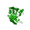
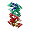
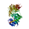
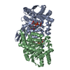
Method to determine structure: MAD / Resolution: 1.9→41.01 Å / Cor.coef. Fo:Fc: 0.956 / Cor.coef. Fo:Fc free: 0.941 / SU B: 6.093 / SU ML: 0.088 / TLS residual ADP flag: LIKELY RESIDUAL / Cross valid method: THROUGHOUT / ESU R: 0.129 / ESU R Free: 0.122 / Stereochemistry target values: MAXIMUM LIKELIHOOD Details: 1. HYDROGENS HAVE BEEN ADDED IN THE RIDING POSITIONS 2. THE DENSITY FOR LOOP 78-83,330-335 ARE POOR, MODEL FOR THESE TWO LOOPS ARE LESS RELIABLE 3. BIOLOGICAL UNIT AS A DIMER IS INFERRED ...Details: 1. HYDROGENS HAVE BEEN ADDED IN THE RIDING POSITIONS 2. THE DENSITY FOR LOOP 78-83,330-335 ARE POOR, MODEL FOR THESE TWO LOOPS ARE LESS RELIABLE 3. BIOLOGICAL UNIT AS A DIMER IS INFERRED FROM HOMOLOGOUS PROTEINS (1A05,1CM7)
Rfactor
Num. reflection
% reflection
Selection details
Rfree
0.21822
1801
5 %
RANDOM
Rwork
0.18779
-
-
-
obs
0.18934
34079
98.15 %
-
Solvent computation
Ion probe radii: 0.8 Å / Shrinkage radii: 0.8 Å / VDW probe radii: 1.2 Å / Solvent model: BABINET MODEL WITH MASK
Displacement parameters
Biso mean: 30.46 Å2
Baniso -1
Baniso -2
Baniso -3
1-
1.45 Å2
0.73 Å2
0 Å2
2-
-
1.45 Å2
0 Å2
3-
-
-
-2.18 Å2
Refinement step
Cycle: LAST / Resolution: 1.9→41.01 Å
Protein
Nucleic acid
Ligand
Solvent
Total
Num. atoms
2723
0
1
106
2830
Refine LS restraints
Refine-ID
Type
Dev ideal
Dev ideal target
Number
X-RAY DIFFRACTION
r_bond_refined_d
0.018
0.022
2785
X-RAY DIFFRACTION
r_bond_other_d
0.002
0.02
2647
X-RAY DIFFRACTION
r_angle_refined_deg
1.673
1.959
3775
X-RAY DIFFRACTION
r_angle_other_deg
0.872
3
6101
X-RAY DIFFRACTION
r_dihedral_angle_1_deg
6.197
5
361
X-RAY DIFFRACTION
r_dihedral_angle_2_deg
31.297
23.333
105
X-RAY DIFFRACTION
r_dihedral_angle_3_deg
13.779
15
472
X-RAY DIFFRACTION
r_dihedral_angle_4_deg
19.243
15
18
X-RAY DIFFRACTION
r_chiral_restr
0.109
0.2
435
X-RAY DIFFRACTION
r_gen_planes_refined
0.007
0.02
3092
X-RAY DIFFRACTION
r_gen_planes_other
0.001
0.02
543
X-RAY DIFFRACTION
r_nbd_refined
0.211
0.2
556
X-RAY DIFFRACTION
r_nbd_other
0.178
0.2
2660
X-RAY DIFFRACTION
r_nbtor_other
0.087
0.2
1671
X-RAY DIFFRACTION
r_xyhbond_nbd_refined
0.164
0.2
99
X-RAY DIFFRACTION
r_symmetry_vdw_refined
0.146
0.2
18
X-RAY DIFFRACTION
r_symmetry_vdw_other
0.219
0.2
65
X-RAY DIFFRACTION
r_symmetry_hbond_refined
0.129
0.2
3
X-RAY DIFFRACTION
r_mcbond_it
2.902
3
1845
X-RAY DIFFRACTION
r_mcbond_other
0.748
3
736
X-RAY DIFFRACTION
r_mcangle_it
3.795
5
2882
X-RAY DIFFRACTION
r_scbond_it
6.495
8
1061
X-RAY DIFFRACTION
r_scangle_it
8.908
11
893
X-RAY DIFFRACTION
r_nbtor_refined
0.178
0.2
1392
LS refinement shell
Resolution: 1.9→1.949 Å / Total num. of bins used: 20
Rfactor
Num. reflection
% reflection
Rfree
0.262
125
5.36 %
Rwork
0.229
2207
-
obs
-
-
87.67 %
Refinement TLS params.
Method: refined / Origin x: 18.1169 Å / Origin y: 86.0493 Å / Origin z: 21.2791 Å
11
12
13
21
22
23
31
32
33
T
-0.0666 Å2
-0.0005 Å2
0.0279 Å2
-
-0.0592 Å2
0.0235 Å2
-
-
0.0526 Å2
L
0.3492 °2
-0.3432 °2
-0.0573 °2
-
2.2755 °2
0.4393 °2
-
-
0.3667 °2
S
-0.0144 Å °
-0.0637 Å °
0.0768 Å °
-0.0791 Å °
0.0112 Å °
-0.3824 Å °
0.0599 Å °
0.0158 Å °
0.0032 Å °
Refinement TLS group
Selection: ALL
+
About Yorodumi
-
News
-
Feb 9, 2022. New format data for meta-information of EMDB entries
New format data for meta-information of EMDB entries
Version 3 of the EMDB header file is now the official format.
The previous official version 1.9 will be removed from the archive.
In the structure databanks used in Yorodumi, some data are registered as the other names, "COVID-19 virus" and "2019-nCoV". Here are the details of the virus and the list of structure data.
Jan 31, 2019. EMDB accession codes are about to change! (news from PDBe EMDB page)
EMDB accession codes are about to change! (news from PDBe EMDB page)
The allocation of 4 digits for EMDB accession codes will soon come to an end. Whilst these codes will remain in use, new EMDB accession codes will include an additional digit and will expand incrementally as the available range of codes is exhausted. The current 4-digit format prefixed with “EMD-” (i.e. EMD-XXXX) will advance to a 5-digit format (i.e. EMD-XXXXX), and so on. It is currently estimated that the 4-digit codes will be depleted around Spring 2019, at which point the 5-digit format will come into force.
The EM Navigator/Yorodumi systems omit the EMD- prefix.
Related info.:Q: What is EMD? / ID/Accession-code notation in Yorodumi/EM Navigator
Yorodumi is a browser for structure data from EMDB, PDB, SASBDB, etc.
This page is also the successor to EM Navigator detail page, and also detail information page/front-end page for Omokage search.
The word "yorodu" (or yorozu) is an old Japanese word meaning "ten thousand". "mi" (miru) is to see.
Related info.:EMDB / PDB / SASBDB / Comparison of 3 databanks / Yorodumi Search / Aug 31, 2016. New EM Navigator & Yorodumi / Yorodumi Papers / Jmol/JSmol / Function and homology information / Changes in new EM Navigator and Yorodumi
 Movie
Movie Controller

 Yorodumi
 Open data
 Basic information
 Components
 Keywords
 Function and homology information

 Authors
 Citation
 Structure visualization
 Downloads & links
 Other downloads

 PDBj
PDBj

 Assembly


 Mass: 35.453 Da / Num. of mol.: 1 / Source method: obtained synthetically / Formula: Cl
 Mass: 18.015 Da / Num. of mol.: 106 / Source method: isolated from a natural source / Formula: H2O
 Sample preparation
 Processing