 Movie
Movie Controller

[English] 日本語
 Yorodumi
- PDB-1vjh: Crystal structure of gene product of At1g24000 from Arabidopsis t... -
+ Open data

- Basic information
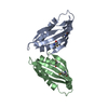
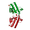
| Entry | Database: PDB / ID: 1vjh | ||||||
|---|---|---|---|---|---|---|---|
| Title | Crystal structure of gene product of At1g24000 from Arabidopsis thaliana | ||||||
 Components Components | Bet v I allergen family | ||||||
 Keywords Keywords |  PLANT PROTEIN / Structural genomics / Arabidopsis Thaliana / Center for Eukaryotic Structural Genomics / Protein Structure Initiative / CESG PLANT PROTEIN / Structural genomics / Arabidopsis Thaliana / Center for Eukaryotic Structural Genomics / Protein Structure Initiative / CESG | ||||||
| Function / homology |  Function and homology informationabscisic acid binding / abscisic acid-activated signaling pathway / protein phosphatase inhibitor activity / defense response / signaling receptor activity / nucleus / cytosol Function and homology informationabscisic acid binding / abscisic acid-activated signaling pathway / protein phosphatase inhibitor activity / defense response / signaling receptor activity / nucleus / cytosolSimilarity search - Function | ||||||
| Biological species |  Arabidopsis thaliana (thale cress) Arabidopsis thaliana (thale cress) | ||||||
| Method | X-RAY DIFFRACTION / SYNCHROTRON / SAD / Resolution: 2.1 Å | ||||||
 Authors Authors | Wesenberg, G.E. / Smith, D.W. / Phillips Jr., G.N. / Johnson, K.A. / Bingman, C.A. / Center for Eukaryotic Structural Genomics (CESG) | ||||||
 Citation Citation | Journal: J.Biomol.Nmr / Year: 2005 Title: 1H, 15N and 13C resonance assignments of the putative Bet v 1 family protein At1g24000.1 from Arabidopsis thaliana. Authors: Song, J. / Zhao, Q. / Lee, M.S. / Markley, J.L. | ||||||
| History |
| ||||||
| Remark 300 | BIOMOLECULE: THIS ENTRY CONTAINS THE CRYSTALLOGRAPHIC ASYMMETRIC UNIT WHICH CONSISTS OF 2 CHAIN(S). ...BIOMOLECULE: THIS ENTRY CONTAINS THE CRYSTALLOGRAPHIC ASYMMETRIC UNIT WHICH CONSISTS OF 2 CHAIN(S). THE AUTHORS ARE UNCERTAIN OF THE BIOLOGICAL UNIT. |
- Structure visualization
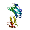
| Structure viewer | Molecule: MolmilJmol/JSmol |
|---|
- Downloads & links
-Download
| PDBx/mmCIF format | 1vjh.cif.gz | 62.4 KB | Display | PDBx/mmCIF format |
|---|---|---|---|---|
| PDB format | pdb1vjh.ent.gz | 45.4 KB | Display | PDB format |
| PDBx/mmJSON format | 1vjh.json.gz | Tree view | PDBx/mmJSON format | |
| Others |  Other downloads Other downloads |
-Validation report
| Arichive directory | https://data.pdbj.org/pub/pdb/validation_reports/vj/1vjhftp://data.pdbj.org/pub/pdb/validation_reports/vj/1vjh | HTTPS FTP |
|---|
-Related structure data
| Similar structure data | |
|---|---|
| Other databases |


-Links
 PDBj
PDBj- Assembly
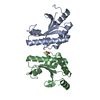
| Deposited unit | 
| ||||||||||
|---|---|---|---|---|---|---|---|---|---|---|---|
| 1 |
| ||||||||||
| Unit cell |
| ||||||||||
| Components on special symmetry positions |
|
-Components
| #1: Protein | Mass: 13873.567 Da / Num. of mol.: 2 Source method: isolated from a genetically manipulated source Source: (gene. exp.) Arabidopsis thaliana (thale cress) / Gene: At1g24000 / Production host:  Escherichia coli (E. coli) / References: UniProt: Q9LR93, UniProt: P0C0B0*PLUS Escherichia coli (E. coli) / References: UniProt: Q9LR93, UniProt: P0C0B0*PLUS#2: Water | ChemComp-HOH / | Water Mass: 18.015 Da / Num. of mol.: 106 / Source method: isolated from a natural source / Formula: H2O Mass: 18.015 Da / Num. of mol.: 106 / Source method: isolated from a natural source / Formula: H2O |
|---|
-Experimental details
-Experiment
| Experiment | Method: X-RAY DIFFRACTION |
|---|
- Sample preparation
| Crystal | Density Matthews: 2.22 Å3/Da / Density % sol: 44.49 % |
|---|---|
| Crystal grow | Temperature: 295 K / Method: hanging drop / pH: 7.5 Details: Sodium Malonate, Sodium HEPES, DMEPEG 550, pH 7.5, VAPOR DIFFUSION, HANGING DROP, temperature 295K, hanging drop |
| Crystal grow | *PLUS Method: unknownDetails: Aceti, D.J., (2003) Biopolymers, 469., Jeon, W.B., (2005) J.Struct.Funct.Genom., 61, 206., Zhao, Q., (2004) J.Struct.Funct.Genom., 5, 87. |
-Data collection
| Diffraction source | Source: SYNCHROTRON / Site: APS  / Beamline: 32-ID / Wavelength: 0.97921 Å / Beamline: 32-ID / Wavelength: 0.97921 Å | |||||||||||||||||||||||||||||||||||||||||||||||||||||||||||||||
|---|---|---|---|---|---|---|---|---|---|---|---|---|---|---|---|---|---|---|---|---|---|---|---|---|---|---|---|---|---|---|---|---|---|---|---|---|---|---|---|---|---|---|---|---|---|---|---|---|---|---|---|---|---|---|---|---|---|---|---|---|---|---|---|---|
| Detector | Type: MARRESEARCH / Detector: CCD / Date: Dec 1, 2003 | |||||||||||||||||||||||||||||||||||||||||||||||||||||||||||||||
| Radiation | Protocol: SINGLE WAVELENGTH / Monochromatic (M) / Laue (L): M / Scattering type: x-ray | |||||||||||||||||||||||||||||||||||||||||||||||||||||||||||||||
| Radiation wavelength | Wavelength: 0.97921 Å / Relative weight: 1 | |||||||||||||||||||||||||||||||||||||||||||||||||||||||||||||||
| Reflection | Resolution: 2.1→30 Å / Num. obs: 14582 / % possible obs: 99.8 % / Rmerge(I) obs: 0.065 | |||||||||||||||||||||||||||||||||||||||||||||||||||||||||||||||
| Reflection shell |
|
-Phasing
| Phasing | Method: SAD | |||||||||||||||||||||||||||||||||||||||||||||||||
|---|---|---|---|---|---|---|---|---|---|---|---|---|---|---|---|---|---|---|---|---|---|---|---|---|---|---|---|---|---|---|---|---|---|---|---|---|---|---|---|---|---|---|---|---|---|---|---|---|---|---|
| Phasing MAD | D res high: 2.1 Å / D res low: 30 Å / FOM : 0.45 / Reflection: 14224 | |||||||||||||||||||||||||||||||||||||||||||||||||
| Phasing MAD set site |
| |||||||||||||||||||||||||||||||||||||||||||||||||
| Phasing MAD shell |
| |||||||||||||||||||||||||||||||||||||||||||||||||
| Phasing dm | FOM : 0.66 / FOM acentric: 0.67 / FOM centric: 0.56 / Reflection: 14229 / Reflection acentric: 13044 / Reflection centric: 1185 | |||||||||||||||||||||||||||||||||||||||||||||||||
| Phasing dm shell |
|
- Processing
| Software |
| ||||||||||||||||||||||||||||||||||||||||||||||||||||||||||||||||||||||||||||||||||||||||||||||||||||||||||||||||||||||||
|---|---|---|---|---|---|---|---|---|---|---|---|---|---|---|---|---|---|---|---|---|---|---|---|---|---|---|---|---|---|---|---|---|---|---|---|---|---|---|---|---|---|---|---|---|---|---|---|---|---|---|---|---|---|---|---|---|---|---|---|---|---|---|---|---|---|---|---|---|---|---|---|---|---|---|---|---|---|---|---|---|---|---|---|---|---|---|---|---|---|---|---|---|---|---|---|---|---|---|---|---|---|---|---|---|---|---|---|---|---|---|---|---|---|---|---|---|---|---|---|---|---|
| Refinement | Resolution: 2.1→21.93 Å / Cor.coef. Fo:Fc: 0.939 / Cor.coef. Fo:Fc free: 0.912 / SU B: 4.033 / SU ML: 0.114 / SU R Cruickshank DPI: 0.234 / Cross valid method: THROUGHOUT / ESU R Free: 0.195
| ||||||||||||||||||||||||||||||||||||||||||||||||||||||||||||||||||||||||||||||||||||||||||||||||||||||||||||||||||||||||
| Solvent computation | Ion probe radii: 0.8 Å / Shrinkage radii: 0.8 Å / VDW probe radii: 1.4 Å / Solvent model: BABINET MODEL PLUS MASK | ||||||||||||||||||||||||||||||||||||||||||||||||||||||||||||||||||||||||||||||||||||||||||||||||||||||||||||||||||||||||
| Displacement parameters | Biso mean: 24.432 Å2
| ||||||||||||||||||||||||||||||||||||||||||||||||||||||||||||||||||||||||||||||||||||||||||||||||||||||||||||||||||||||||
| Refinement step | Cycle: LAST / Resolution: 2.1→21.93 Å
| ||||||||||||||||||||||||||||||||||||||||||||||||||||||||||||||||||||||||||||||||||||||||||||||||||||||||||||||||||||||||
| Refine LS restraints |
| ||||||||||||||||||||||||||||||||||||||||||||||||||||||||||||||||||||||||||||||||||||||||||||||||||||||||||||||||||||||||
| LS refinement shell | Refine-ID: X-RAY DIFFRACTION / Total num. of bins used: 19
| ||||||||||||||||||||||||||||||||||||||||||||||||||||||||||||||||||||||||||||||||||||||||||||||||||||||||||||||||||||||||
| Software | *PLUS Name: REFMAC / Version: 5.1.24 / Classification: refinement | ||||||||||||||||||||||||||||||||||||||||||||||||||||||||||||||||||||||||||||||||||||||||||||||||||||||||||||||||||||||||
| Refine LS restraints | *PLUS
|
