 Movie
Movie Controller


+ Open data

- Basic information
| Entry | Database: PDB / ID: 1vgq | ||||||
|---|---|---|---|---|---|---|---|
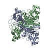
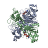
| Title | Formyl-CoA transferase mutant Asp169 to Ala | ||||||
 Components Components | Formyl-coenzyme A transferase | ||||||
 Keywords Keywords |  TRANSFERASE / CoA-transferase / oxalate / oxalate degradation / intertwined / knotted fold / CAIB-BAIF family / CoA complex TRANSFERASE / CoA-transferase / oxalate / oxalate degradation / intertwined / knotted fold / CAIB-BAIF family / CoA complex | ||||||
| Function / homology |  Function and homology informationformyl-CoA transferase / formyl-CoA transferase activity / oxalate catabolic process / cytoplasm Function and homology informationformyl-CoA transferase / formyl-CoA transferase activity / oxalate catabolic process / cytoplasmSimilarity search - Function | ||||||
| Biological species |  Oxalobacter formigenes (bacteria) Oxalobacter formigenes (bacteria) | ||||||
| Method | X-RAY DIFFRACTION / SYNCHROTRON / FOURIER SYNTHESIS / Resolution: 2.13 Å | ||||||
 Authors Authors | Ricagno, S. / Jonsson, S. / Richards, N.G. / Lindqvist, Y. | ||||||
 Citation Citation | Journal: J.Biol.Chem. / Year: 2004 Title: Kinetic and mechanistic characterization of the formyl-CoA transferase from Oxalobacter formigenes Authors: Jonsson, S. / Ricagno, S. / Lindqvist, Y. / Richards, N.G. | ||||||
| History |
|
- Structure visualization
| Structure viewer | Molecule: MolmilJmol/JSmol |
|---|
- Downloads & links
-Download
| PDBx/mmCIF format | 1vgq.cif.gz | 185.6 KB | Display | PDBx/mmCIF format |
|---|---|---|---|---|
| PDB format | pdb1vgq.ent.gz | 147.6 KB | Display | PDB format |
| PDBx/mmJSON format | 1vgq.json.gz | Tree view | PDBx/mmJSON format | |
| Others |  Other downloads Other downloads |
-Validation report
| Arichive directory | https://data.pdbj.org/pub/pdb/validation_reports/vg/1vgqftp://data.pdbj.org/pub/pdb/validation_reports/vg/1vgq | HTTPS FTP |
|---|
-Related structure data
| Related structure data | 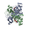 1t3zC  1t4cC 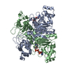 1vgrC  1p5rS S: Starting model for refinement C: citing same article ( |
|---|---|
| Similar structure data |


-Links
 PDBj
PDBj- Assembly
| Deposited unit | 
| ||||||||
|---|---|---|---|---|---|---|---|---|---|
| 1 |
| ||||||||
| Unit cell |
|
-Components
| #1: Protein | Mass: 47187.656 Da / Num. of mol.: 2 / Mutation: D169A Source method: isolated from a genetically manipulated source Source: (gene. exp.) Oxalobacter formigenes (bacteria) / Gene: FRC / Plasmid: pET9a / Species (production host): Escherichia coli / Production host: Escherichia coli BL21(DE3) (bacteria) / Strain (production host): BL21 (DE3) / References: UniProt: O06644, formyl-CoA transferase#2: Chemical |   Mass: 783.534 Da / Num. of mol.: 2 / Source method: obtained synthetically / Formula: C21H36N7O17P3S Mass: 783.534 Da / Num. of mol.: 2 / Source method: obtained synthetically / Formula: C21H36N7O17P3S#3: Water | ChemComp-HOH / | Water Mass: 18.015 Da / Num. of mol.: 470 / Source method: isolated from a natural source / Formula: H2O Mass: 18.015 Da / Num. of mol.: 470 / Source method: isolated from a natural source / Formula: H2O |
|---|
-Experimental details
-Experiment
| Experiment | Method: X-RAY DIFFRACTION / Number of used crystals: 1 |
|---|
- Sample preparation
| Crystal | Density Matthews: 2.82 Å3/Da / Density % sol: 56.1 % |
|---|---|
| Crystal grow | Temperature: 293 K / Method: vapor diffusion, hanging drop / pH: 7.5 Details: PEG 4000, Mg chloride, Hepes, pH 7.5, VAPOR DIFFUSION, HANGING DROP, temperature 293K |
-Data collection
| Diffraction | Mean temperature: 110 K |
|---|---|
| Diffraction source | Source: SYNCHROTRON / Site: EMBL/DESY, HAMBURG  / Beamline: X11 / Wavelength: 0.812 Å / Beamline: X11 / Wavelength: 0.812 Å |
| Detector | Type: MARRESEARCH / Detector: CCD / Date: Sep 2, 2003 |
| Radiation | Protocol: SINGLE WAVELENGTH / Monochromatic (M) / Laue (L): M / Scattering type: x-ray |
| Radiation wavelength | Wavelength: 0.812 Å / Relative weight: 1 |
| Reflection | Resolution: 2.13→20.17 Å / Num. all: 61071 / Num. obs: 61071 / % possible obs: 98.2 % / Observed criterion σ(F): 0 / Observed criterion σ(I): 0 / Redundancy: 4.3 % / Biso Wilson estimate: 31.3 Å2 / Rsym value: 0.091 / Net I/σ(I): 13.7 |
| Reflection shell | Resolution: 2.13→2.24 Å / Redundancy: 3.3 % / Mean I/σ(I) obs: 2.2 / Num. unique all: 7477 / Rsym value: 0.417 / % possible all: 81.9 |
- Processing
| Software |
| ||||||||||||||||||||||||||||||||||||||||||||||||||||||||||||||||||||||||||||||||||||||||||||||||||||
|---|---|---|---|---|---|---|---|---|---|---|---|---|---|---|---|---|---|---|---|---|---|---|---|---|---|---|---|---|---|---|---|---|---|---|---|---|---|---|---|---|---|---|---|---|---|---|---|---|---|---|---|---|---|---|---|---|---|---|---|---|---|---|---|---|---|---|---|---|---|---|---|---|---|---|---|---|---|---|---|---|---|---|---|---|---|---|---|---|---|---|---|---|---|---|---|---|---|---|---|---|---|
| Refinement | Method to determine structure: FOURIER SYNTHESIS Starting model: PDB ENTRY 1P5R Resolution: 2.13→20.17 Å / Cor.coef. Fo:Fc: 0.959 / Cor.coef. Fo:Fc free: 0.944 / SU B: 3.992 / SU ML: 0.103 / TLS residual ADP flag: LIKELY RESIDUAL / Cross valid method: THROUGHOUT / σ(F): 0 / σ(I): 0 / ESU R: 0.185 / ESU R Free: 0.155 / Stereochemistry target values: MAXIMUM LIKELIHOOD / Details: HYDROGENS HAVE BEEN ADDED IN THE RIDING POSITIONS
| ||||||||||||||||||||||||||||||||||||||||||||||||||||||||||||||||||||||||||||||||||||||||||||||||||||
| Solvent computation | Ion probe radii: 0.8 Å / Shrinkage radii: 0.8 Å / VDW probe radii: 1.4 Å / Solvent model: BABINET MODEL WITH MASK | ||||||||||||||||||||||||||||||||||||||||||||||||||||||||||||||||||||||||||||||||||||||||||||||||||||
| Displacement parameters | Biso mean: 30.873 Å2
| ||||||||||||||||||||||||||||||||||||||||||||||||||||||||||||||||||||||||||||||||||||||||||||||||||||
| Refinement step | Cycle: LAST / Resolution: 2.13→20.17 Å
| ||||||||||||||||||||||||||||||||||||||||||||||||||||||||||||||||||||||||||||||||||||||||||||||||||||
| Refine LS restraints |
| ||||||||||||||||||||||||||||||||||||||||||||||||||||||||||||||||||||||||||||||||||||||||||||||||||||
| LS refinement shell | Resolution: 2.135→2.19 Å / Total num. of bins used: 20 /
| ||||||||||||||||||||||||||||||||||||||||||||||||||||||||||||||||||||||||||||||||||||||||||||||||||||
| Refinement TLS params. | Method: refined / Refine-ID: X-RAY DIFFRACTION
| ||||||||||||||||||||||||||||||||||||||||||||||||||||||||||||||||||||||||||||||||||||||||||||||||||||
| Refinement TLS group |
|
