 Movie
Movie Controller


+ Open data

- Basic information
| Entry | Database: PDB / ID: 1veu | ||||||
|---|---|---|---|---|---|---|---|
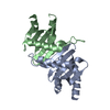
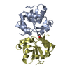
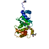
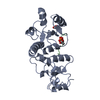
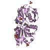
| Title | Crystal structure of the p14/MP1 complex at 2.15 A resolution | ||||||
 Components Components |
| ||||||
 Keywords Keywords |  SIGNALING PROTEIN/PROTEIN BINDING / profilin / scaffold / adaptor / SIGNALING PROTEIN-PROTEIN BINDING COMPLEX SIGNALING PROTEIN/PROTEIN BINDING / profilin / scaffold / adaptor / SIGNALING PROTEIN-PROTEIN BINDING COMPLEX | ||||||
| Function / homology |  Function and homology information Function and homology informationEnergy dependent regulation of mTOR by LKB1-AMPK / Amino acids regulate mTORC1 / MTOR signalling / Regulation of PTEN gene transcription / Macroautophagy / regulation of cell-substrate junction organization / TP53 Regulates Metabolic Genes / mTORC1-mediated signalling / FNIP-folliculin RagC/D GAP / Ragulator complex ...Energy dependent regulation of mTOR by LKB1-AMPK / Amino acids regulate mTORC1 / MTOR signalling / Regulation of PTEN gene transcription / Macroautophagy / regulation of cell-substrate junction organization / TP53 Regulates Metabolic Genes / mTORC1-mediated signalling / FNIP-folliculin RagC/D GAP / Ragulator complex / MAP2K and MAPK activation / protein localization to cell junction / TORC1 signaling / fibroblast migration / kinase activator activity / positive regulation of TOR signaling / positive regulation of TORC1 signaling / Neutrophil degranulation / guanyl-nucleotide exchange factor activity / regulation of cell growth / cellular response to amino acid stimulus / protein localization / late endosome / late endosome membrane / positive regulation of MAPK cascade / molecular adaptor activity / lysosomal membraneSimilarity search - Function | ||||||
| Biological species |  Mus musculus (house mouse) Mus musculus (house mouse) | ||||||
| Method | X-RAY DIFFRACTION / MOLECULAR REPLACEMENT / Resolution: 2.15 Å | ||||||
 Authors Authors | Kurzbauer, R. / Teis, D. / Maurer-Stroh, S. / Eisenhaber, F. / Hekman, M. / Bourenkov, G.P. / Bartunik, H.D. / Huber, L.A. / Clausen, T. | ||||||
 Citation Citation | Journal: Proc.Natl.Acad.Sci.USA / Year: 2004 Title: Crystal structure of the p14/MP1 scaffolding complex: How a twin couple attaches mitogen- activated protein kinase signaling to late endosomes Authors: Kurzbauer, R. / Teis, D. / De Araujo, M.E. / Maurer-Stroh, S. / Eisenhaber, F. / Bourenkov, G.P. / Bartunik, H.D. / Hekman, M. / Rapp, U.R. / Huber, L.A. / Clausen, T. | ||||||
| History |
|
- Structure visualization
| Structure viewer | Molecule: MolmilJmol/JSmol |
|---|
- Downloads & links
-Download
| PDBx/mmCIF format | 1veu.cif.gz | 60.7 KB | Display | PDBx/mmCIF format |
|---|---|---|---|---|
| PDB format | pdb1veu.ent.gz | 43.7 KB | Display | PDB format |
| PDBx/mmJSON format | 1veu.json.gz | Tree view | PDBx/mmJSON format | |
| Others |  Other downloads Other downloads |
-Validation report
| Arichive directory | https://data.pdbj.org/pub/pdb/validation_reports/ve/1veuftp://data.pdbj.org/pub/pdb/validation_reports/ve/1veu | HTTPS FTP |
|---|
-Related structure data
| Related structure data | 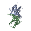 1vetSC S: Starting model for refinement C: citing same article ( |
|---|---|
| Similar structure data |


-Links
 PDBj
PDBj


- Assembly
| Deposited unit | 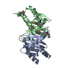
| ||||||||
|---|---|---|---|---|---|---|---|---|---|
| 1 |
| ||||||||
| Unit cell |
|
-Components
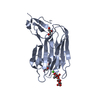
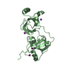
| #1: Protein | Mass: 13614.563 Da / Num. of mol.: 1 Source method: isolated from a genetically manipulated source Source: (gene. exp.) Mus musculus (house mouse) / Production host:  Escherichia coli (E. coli) / References: UniProt: O88653 Escherichia coli (E. coli) / References: UniProt: O88653 |
|---|---|
| #2: Protein | Mass: 13781.953 Da / Num. of mol.: 1 Source method: isolated from a genetically manipulated source Source: (gene. exp.) Mus musculus (house mouse) / Production host: Escherichia coli (E. coli) / References: UniProt: Q9JHS3 |
| #3: Water | ChemComp-HOH / Water Mass: 18.015 Da / Num. of mol.: 83 / Source method: isolated from a natural source / Formula: H2O Mass: 18.015 Da / Num. of mol.: 83 / Source method: isolated from a natural source / Formula: H2O |
-Experimental details
-Experiment
| Experiment | Method: X-RAY DIFFRACTION / Number of used crystals: 1 |
|---|
- Sample preparation
| Crystal | Density Matthews: 1.6 Å3/Da / Density % sol: 33 % |
|---|---|
| Crystal grow | Temperature: 292 K / Method: vapor diffusion, sitting drop / pH: 8.5 Details: PEG4000, Tris, Magnesium Chloride, Sodium/Potassium Phosphate, pH 8.5, VAPOR DIFFUSION, SITTING DROP, temperature 292K |
-Data collection
| Diffraction | Mean temperature: 100 K |
|---|---|
| Diffraction source | Source: ROTATING ANODE / Type: RIGAKU RU300 / Wavelength: 1.5418 Å |
| Detector | Type: MARRESEARCH / Detector: IMAGE PLATE / Date: Jan 16, 2004 |
| Radiation | Monochromator: filter / Protocol: SINGLE WAVELENGTH / Monochromatic (M) / Laue (L): M / Scattering type: x-ray |
| Radiation wavelength | Wavelength: 1.5418 Å / Relative weight: 1 |
| Reflection | Resolution: 2.15→20 Å / Num. all: 33808 / Num. obs: 11596 / % possible obs: 94.1 % / Observed criterion σ(F): 0 / Observed criterion σ(I): 0 |
| Reflection shell | Resolution: 2.15→2.23 Å / % possible all: 92.5 |
- Processing
| Software |
| ||||||||||||||||||||
|---|---|---|---|---|---|---|---|---|---|---|---|---|---|---|---|---|---|---|---|---|---|
| Refinement | Method to determine structure: MOLECULAR REPLACEMENT Starting model: 1vet Resolution: 2.15→20 Å / σ(F): 0 / σ(I): 0 / Stereochemistry target values: Engh & Huber
| ||||||||||||||||||||
| Refinement step | Cycle: LAST / Resolution: 2.15→20 Å
| ||||||||||||||||||||
| Refine LS restraints |
|
