 Movie
Movie Controller

[English] 日本語
 Yorodumi
- PDB-1vea: Crystal Structure of HutP, an RNA binding antitermination protein -
+ Open data

- Basic information
| Entry | Database: PDB / ID: 1vea | ||||||
|---|---|---|---|---|---|---|---|
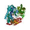
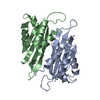
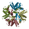
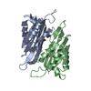
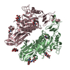
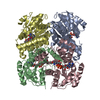
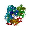
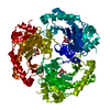
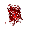
| Title | Crystal Structure of HutP, an RNA binding antitermination protein | ||||||
 Components Components | Hut operon positive regulatory protein | ||||||
 Keywords Keywords |  RNA BINDING PROTEIN / HutP / Antiterminator / regulation of transcription RNA BINDING PROTEIN / HutP / Antiterminator / regulation of transcription | ||||||
| Function / homology |  Function and homology information Function and homology informationL-histidine metabolic process / mRNA binding / positive regulation of gene expressionSimilarity search - Function | ||||||
| Biological species |  Bacillus subtilis (bacteria) Bacillus subtilis (bacteria) | ||||||
| Method | X-RAY DIFFRACTION / SYNCHROTRON / MAD / Resolution: 2.8 Å | ||||||
 Authors Authors | Kumarevel, T.S. / Fujimoto, Z. / Karthe, P. / Oda, M. / Mizuno, H. / Kumar, P.K.R. | ||||||
 Citation Citation | Journal: Structure / Year: 2004 Title: Crystal Structure of Activated HutP; An RNA Binding Protein that Regulates Transcription of the hut Operon in Bacillus subtilis Authors: Kumarevel, T.S. / Fujimoto, Z. / Karthe, P. / Oda, M. / Mizuno, H. / Kumar, P.K.R. #1: Journal: J.Struct.Biol. / Year: 2002 Title: Crystallization and preliminary X-ray diffraction studies of HutP protein: an RNA-binding protein that regulates the transcription of hut operon in Bacillus subtilis Authors: Kumarevel, T.S. / Fujimoto, Z. / Padmanabhan, B. / Oda, M. / Nishikawa, S. / Mizuno, H. / Kumar, P.K.R. #2: Journal: Mol.Microbiol. / Year: 2000 Title: cis-acting regulatory sequences for antitermination in the transcript of the Bacillus subtilis hut operon and histidine-dependent binding of HutP to the transcript containing the regulatory sequences Authors: Oda, M. / Kobayashi, N. / Ito, A. / Kurusu, Y. / Taira, K. | ||||||
| History |
|
- Structure visualization
| Structure viewer | Molecule: MolmilJmol/JSmol |
|---|
- Downloads & links
-Download
| PDBx/mmCIF format | 1vea.cif.gz | 68.7 KB | Display | PDBx/mmCIF format |
|---|---|---|---|---|
| PDB format | pdb1vea.ent.gz | 51.3 KB | Display | PDB format |
| PDBx/mmJSON format | 1vea.json.gz | Tree view | PDBx/mmJSON format | |
| Others |  Other downloads Other downloads |
-Validation report
| Arichive directory | https://data.pdbj.org/pub/pdb/validation_reports/ve/1veaftp://data.pdbj.org/pub/pdb/validation_reports/ve/1vea | HTTPS FTP |
|---|
-Related structure data
| Similar structure data |
|---|

-Links
 PDBj
PDBj- Assembly
| Deposited unit | 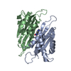
| ||||||||
|---|---|---|---|---|---|---|---|---|---|
| 1 | 
| ||||||||
| Unit cell |
| ||||||||
| Details | The biological assembly is a hexamer generated from the dimer in the asymmetric unit by the operations: z,x,y and y,z,x. |
-Components
| #1: Protein | Mass: 16232.553 Da / Num. of mol.: 2 / Fragment: RNA-binding, antiterminator protein / Mutation: V51I Source method: isolated from a genetically manipulated source Source: (gene. exp.) Bacillus subtilis (bacteria) / Plasmid: pET5a, pETHP4 / Species (production host): Escherichia coli / Production host: Escherichia coli BL21(DE3) (bacteria) / Strain (production host): BL21(DE3) / References: UniProt: P10943#2: Chemical | ChemComp-HBN / |   Type: L-peptide linking / Mass: 280.324 Da / Num. of mol.: 1 / Source method: obtained synthetically / Formula: C16H16N4O Type: L-peptide linking / Mass: 280.324 Da / Num. of mol.: 1 / Source method: obtained synthetically / Formula: C16H16N4O#3: Water | ChemComp-HOH / | Water Mass: 18.015 Da / Num. of mol.: 83 / Source method: isolated from a natural source / Formula: H2O Mass: 18.015 Da / Num. of mol.: 83 / Source method: isolated from a natural source / Formula: H2O |
|---|
-Experimental details
-Experiment
| Experiment | Method: X-RAY DIFFRACTION / Number of used crystals: 1 |
|---|
- Sample preparation
| Crystal | Density Matthews: 2.23 Å3/Da / Density % sol: 44.82 % |
|---|---|
| Crystal grow | Temperature: 293 K / Method: vapor diffusion, hanging drop / pH: 6.8 Details: PEG2000MME, Pottasium bromide, Na Cacodylate, pH 6.8, VAPOR DIFFUSION, HANGING DROP, temperature 293K |
-Data collection
| Diffraction | Mean temperature: 100 K | |||||||||||||||
|---|---|---|---|---|---|---|---|---|---|---|---|---|---|---|---|---|
| Diffraction source | Source: SYNCHROTRON / Site: Photon Factory  / Beamline: BL-6A / Wavelength: 0.978, 1.007, 0.995, 0.950 / Beamline: BL-6A / Wavelength: 0.978, 1.007, 0.995, 0.950 | |||||||||||||||
| Detector | Type: ADSC QUANTUM 4 / Detector: CCD / Date: Oct 3, 2002 | |||||||||||||||
| Radiation | Monochromator: Si 111 CHANNEL / Protocol: MAD / Monochromatic (M) / Laue (L): M / Scattering type: x-ray | |||||||||||||||
| Radiation wavelength |
| |||||||||||||||
| Reflection | Resolution: 2.8→31.803 Å / Num. all: 7375 / Num. obs: 7375 / % possible obs: 100 % / Observed criterion σ(F): 0 / Observed criterion σ(I): 0 / Redundancy: 7.1 % / Biso Wilson estimate: 85.5 Å2 / Rmerge(I) obs: 0.054 / Rsym value: 0.05 / Net I/σ(I): 7.9 | |||||||||||||||
| Reflection shell | Resolution: 2.8→2.95 Å / Redundancy: 7.3 % / Rmerge(I) obs: 0.258 / Mean I/σ(I) obs: 3.2 / Num. unique all: 7379 / Rsym value: 0.239 / % possible all: 100 |
- Processing
| Software |
| ||||||||||||||||||||||||||||||||||||
|---|---|---|---|---|---|---|---|---|---|---|---|---|---|---|---|---|---|---|---|---|---|---|---|---|---|---|---|---|---|---|---|---|---|---|---|---|---|
| Refinement | Method to determine structure: MAD / Resolution: 2.8→19.48 Å / Rfactor Rfree error: 0.014 / Data cutoff high absF: 1236996.06 / Data cutoff low absF: 0 / Isotropic thermal model: RESTRAINED / Cross valid method: THROUGHOUT / σ(F): 0 / Stereochemistry target values: Engh & Huber
| ||||||||||||||||||||||||||||||||||||
| Solvent computation | Solvent model: FLAT MODEL / Bsol: 26.0642 Å2 / ksol: 0.259333 e/Å3 | ||||||||||||||||||||||||||||||||||||
| Displacement parameters | Biso mean: 68.6 Å2
| ||||||||||||||||||||||||||||||||||||
| Refine analyze |
| ||||||||||||||||||||||||||||||||||||
| Refinement step | Cycle: LAST / Resolution: 2.8→19.48 Å
| ||||||||||||||||||||||||||||||||||||
| Refine LS restraints |
| ||||||||||||||||||||||||||||||||||||
| LS refinement shell | Resolution: 2.8→2.97 Å / Rfactor Rfree error: 0.059 / Total num. of bins used: 6
| ||||||||||||||||||||||||||||||||||||
| Xplor file |
|
