[English] 日本語
Yorodumi
- PDB-1vcx: Neutron Crystal Structure of the Wild Type Rubredoxin from Pyroco... -

+
Open data


ID or keywords:

Loading...

-
Basic information

Entry
Database: PDB / ID: 1vcx
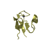
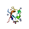
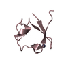
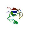
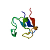
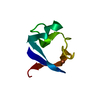
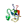
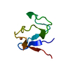
TitleNeutron Crystal Structure of the Wild Type Rubredoxin from Pyrococcus Furiosus at 1.5A Resolution
ComponentsRubredoxin
KeywordsELECTRON TRANSPORT / Neutron structure / Hydrogen atom position / Hydration water structure / Hydrogen/Deuterium exchange / Iron-sulfur core / N-terminal region
Function / homology
Function and homology information


electron transfer activity / iron ion binding
Similarity search - Function
Rubredoxin / Rubredoxin, iron-binding site / Rubredoxin signature. / Rubrerythrin, domain 2 - #10 / Rubredoxin domain / Rubredoxin / Rubredoxin-like domain / Rubredoxin-like domain profile. / Rubrerythrin, domain 2 / Single Sheet / Mainly Beta
Similarity search - Domain/homology
DEUTERATED WATER / : / Rubredoxin
Similarity search - Component
Biological speciesPyrococcus furiosus (archaea)
MethodNEUTRON DIFFRACTION / NUCLEAR REACTOR / MOLECULAR REPLACEMENT / Resolution: 1.5 Å
AuthorsKurihara, K. / Tanaka, I. / Chatake, T. / Adams, M.W.W. / Jenney Jr., F.E. / Moiseeva, N. / Bau, R. / Niimura, N.
CitationJournal: Proc.Natl.Acad.Sci.USA / Year: 2004
Title: Neutron crystallographic study on rubredoxin from Pyrococcus furiosus by BIX-3, a single-crystal diffractometer for biomacromolecules
Authors: Kurihara, K. / Tanaka, I. / Chatake, T. / Adams, M.W.W. / Jenney Jr., F.E. / Moiseeva, N. / Bau, R. / Niimura, N.
History
DepositionMar 17, 2004Deposition site: PDBJ / Processing site: PDBJ
Revision 1.0Aug 10, 2004Provider: repository / Type: Initial release
Revision 1.1Apr 27, 2008Group: Version format compliance
Revision 1.2Jul 13, 2011Group: Non-polymer description / Version format compliance
Revision 1.3Mar 7, 2018Group: Data collection / Category: diffrn_detector / diffrn_radiation
Item: _diffrn_detector.detector / _diffrn_detector.type ..._diffrn_detector.detector / _diffrn_detector.type / _diffrn_radiation.pdbx_diffrn_protocol / _diffrn_radiation.pdbx_monochromatic_or_laue_m_l
Revision 1.4Jul 11, 2018Group: Data collection / Category: diffrn_radiation / diffrn_source
Item: _diffrn_radiation.pdbx_scattering_type / _diffrn_source.pdbx_synchrotron_beamline ..._diffrn_radiation.pdbx_scattering_type / _diffrn_source.pdbx_synchrotron_beamline / _diffrn_source.pdbx_synchrotron_site / _diffrn_source.type
Revision 1.5Jul 18, 2018Group: Data collection / Category: diffrn_radiation
Item: _diffrn_radiation.pdbx_diffrn_protocol / _diffrn_radiation.pdbx_monochromatic_or_laue_m_l
Revision 1.6Oct 25, 2023Group: Data collection / Database references ...Data collection / Database references / Derived calculations / Refinement description
Category: chem_comp_atom / chem_comp_bond ...chem_comp_atom / chem_comp_bond / database_2 / pdbx_initial_refinement_model / struct_conn / struct_conn_type / struct_site
Item: _database_2.pdbx_DOI / _database_2.pdbx_database_accession ..._database_2.pdbx_DOI / _database_2.pdbx_database_accession / _struct_conn.conn_type_id / _struct_conn.id / _struct_conn.pdbx_dist_value / _struct_conn.pdbx_leaving_atom_flag / _struct_conn.ptnr1_auth_comp_id / _struct_conn.ptnr1_auth_seq_id / _struct_conn.ptnr1_label_asym_id / _struct_conn.ptnr1_label_atom_id / _struct_conn.ptnr1_label_comp_id / _struct_conn.ptnr1_label_seq_id / _struct_conn.ptnr2_auth_comp_id / _struct_conn.ptnr2_auth_seq_id / _struct_conn.ptnr2_label_asym_id / _struct_conn.ptnr2_label_atom_id / _struct_conn.ptnr2_label_comp_id / _struct_conn.ptnr2_label_seq_id / _struct_conn_type.id / _struct_site.pdbx_auth_asym_id / _struct_site.pdbx_auth_comp_id / _struct_site.pdbx_auth_seq_id

-
Structure visualization

Structure viewerMolecule:
MolmilJmol/JSmol

Downloads & links

-
Assembly

Deposited unit
A: Rubredoxin
hetero molecules


Theoretical massNumber of molelcules
Total (without water)5,9562
Polymers5,9011
Non-polymers561
Water66737
1


  • Idetical with deposited unit
  • defined by author
TypeNameSymmetry operationNumber
identity operation1_555x,y,z1
Unit cell
Length a, b, c (Å)34.320, 35.310, 44.230
Angle α, β, γ (deg.)90.00, 90.00, 90.00
Int Tables number19
Space group name H-MP212121

-
Components

#1: Protein Rubredoxin / / PF Rd


Mass: 5900.532 Da / Num. of mol.: 1
Source method: isolated from a genetically manipulated source
Source: (gene. exp.) Pyrococcus furiosus (archaea) / Plasmid: pT7-7 / Species (production host): Escherichia coli / Production host: Escherichia coli BL21(DE3) (bacteria) / Strain (production host): BL21-DE3 / References: UniProt: P24297
#2: Chemical ChemComp-FE / FE (III) ION / Iron


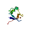
Mass: 55.845 Da / Num. of mol.: 1 / Source method: obtained synthetically / Formula: Fe
#3: Chemical ChemComp-DOD / water / Heavy water


Mass: 18.015 Da / Num. of mol.: 37 / Source method: isolated from a natural source / Formula: D2O

-
Experimental details

-
Experiment

ExperimentMethod: NEUTRON DIFFRACTION / Number of used crystals: 1

-
Sample preparation

Crystal growTemperature: 298 K / Method: macroseeding / pH: 8
Details: sodium dihydrogen phosphate, dipotassium hydrogen phosphate, pH 8.0, Macroseeding, temperature 298K

-
Data collection

DiffractionMean temperature: 298 K
Diffraction sourceSource: NUCLEAR REACTOR / Site: JRR-3M / Beamline: 1G-A / Wavelength: 2.33 Å
DetectorType: MACSCIENCE DIP100 / Detector: IMAGE PLATE / Date: Aug 24, 1999
RadiationMonochromator: elastically-bent perfect Si(111) / Protocol: SINGLE WAVELENGTH / Monochromatic (M) / Laue (L): M / Scattering type: neutron
Radiation wavelengthWavelength: 2.33 Å / Relative weight: 1
ReflectionResolution: 1.5→30 Å / Num. obs: 7417 / % possible obs: 81.9 % / Redundancy: 3.3 % / Biso Wilson estimate: 6.4 Å2 / Rsym value: 0.096 / Net I/σ(I): 8.5
Reflection shellResolution: 1.5→1.55 Å / Redundancy: 1.7 % / Mean I/σ(I) obs: 3.1 / Num. unique all: 469 / Rsym value: 0.232 / % possible all: 53.1

-
Processing

Software
NameVersionClassification
CNS1refinement
DENZOdata reduction
SCALEPACKdata scaling
X-PLORphasing
CNSphasing
RefinementMethod to determine structure: MOLECULAR REPLACEMENT
Starting model: PDB ENTRY 1BRF
Resolution: 1.5→27.6 Å / Rfactor Rfree error: 0.008 / Data cutoff high absF: 103355.67 / Data cutoff low absF: 0 / Isotropic thermal model: RESTRAINED / Cross valid method: THROUGHOUT / σ(F): 2
Stereochemistry target values: MAXIMUM LIKELIHOOD TARGET USING AMPLITUDES
Details: X-PLOR 3.851 was also used for the refinement. THE STANDARD TOPOLOGY AND PARAMETER FILES WERE CHANGED FOR HYDROGEN AND DEUTERIUM ATOM PARAMETERS TO MEET THE REQUIREMENTS OF THE NEUTRON STRUCTURE REFINEMENT.
RfactorNum. reflection% reflectionSelection details
Rfree0.217 789 10.8 %RANDOM
Rwork0.186 ---
obs-7286 80.5 %-
Solvent computationSolvent model: FLAT MODEL / Bsol: 100 Å2 / ksol: 0.05010384 e/Å3
Displacement parametersBiso mean: 11.9 Å2
Baniso -1Baniso -2Baniso -3
1--0.35 Å20 Å20 Å2
2--0.03 Å20 Å2
3---0.32 Å2
Refine analyze
FreeObs
Luzzati coordinate error0.19 Å0.16 Å
Luzzati d res low-5 Å
Luzzati sigma a0.17 Å0.15 Å
Refinement stepCycle: LAST / Resolution: 1.5→27.6 Å
ProteinNucleic acidLigandSolventTotal
Num. atoms413 0 1 37 451
Refine LS restraints
Refine-IDTypeDev idealDev ideal target
NEUTRON DIFFRACTIONc_bond_d0.01
NEUTRON DIFFRACTIONc_angle_deg1.24
NEUTRON DIFFRACTIONc_dihedral_angle_d25.8
NEUTRON DIFFRACTIONc_improper_angle_d1.2
NEUTRON DIFFRACTIONc_mcbond_it1.331.5
NEUTRON DIFFRACTIONc_mcangle_it2.192
NEUTRON DIFFRACTIONc_scbond_it1.892
NEUTRON DIFFRACTIONc_scangle_it2.492.5
LS refinement shellResolution: 1.5→1.55 Å / Rfactor Rfree error: 0.043 / Total num. of bins used: 10
RfactorNum. reflection% reflection
Rfree0.298 48 10.3 %
Rwork0.271 416 -
obs-464 52.5 %
Xplor file
Refine-IDSerial noParam fileTopol file
NEUTRON DIFFRACTION1PROTEIN.PARAMPROTEIN-ALLHDG.TOP
NEUTRON DIFFRACTION2WATER.PARAMWATER.TOP

+
About Yorodumi

-
News

-
Feb 9, 2022. New format data for meta-information of EMDB entries

New format data for meta-information of EMDB entries

  • Version 3 of the EMDB header file is now the official format.
  • The previous official version 1.9 will be removed from the archive.

Related info.:EMDB header

External links:wwPDB to switch to version 3 of the EMDB data model

-
Aug 12, 2020. Covid-19 info

Covid-19 info

URL: https://pdbj.org/emnavi/covid19.php

New page: Covid-19 featured information page in EM Navigator.

Related info.:Covid-19 info / Mar 5, 2020. Novel coronavirus structure data

+
Mar 5, 2020. Novel coronavirus structure data

Novel coronavirus structure data

Related info.:Yorodumi Speices / Aug 12, 2020. Covid-19 info

External links:COVID-19 featured content - PDBj / Molecule of the Month (242):Coronavirus Proteases

+
Jan 31, 2019. EMDB accession codes are about to change! (news from PDBe EMDB page)

EMDB accession codes are about to change! (news from PDBe EMDB page)

  • The allocation of 4 digits for EMDB accession codes will soon come to an end. Whilst these codes will remain in use, new EMDB accession codes will include an additional digit and will expand incrementally as the available range of codes is exhausted. The current 4-digit format prefixed with “EMD-” (i.e. EMD-XXXX) will advance to a 5-digit format (i.e. EMD-XXXXX), and so on. It is currently estimated that the 4-digit codes will be depleted around Spring 2019, at which point the 5-digit format will come into force.
  • The EM Navigator/Yorodumi systems omit the EMD- prefix.

Related info.:Q: What is EMD? / ID/Accession-code notation in Yorodumi/EM Navigator

External links:EMDB Accession Codes are Changing Soon! / Contact to PDBj

+
Jul 12, 2017. Major update of PDB

Major update of PDB

  • wwPDB released updated PDB data conforming to the new PDBx/mmCIF dictionary.
  • This is a major update changing the version number from 4 to 5, and with Remediation, in which all the entries are updated.
  • In this update, many items about electron microscopy experimental information are reorganized (e.g. em_software).
  • Now, EM Navigator and Yorodumi are based on the updated data.

External links:wwPDB Remediation / Enriched Model Files Conforming to OneDep Data Standards Now Available in the PDB FTP Archive

-
Yorodumi

Thousand views of thousand structures

  • Yorodumi is a browser for structure data from EMDB, PDB, SASBDB, etc.
  • This page is also the successor to EM Navigator detail page, and also detail information page/front-end page for Omokage search.
  • The word "yorodu" (or yorozu) is an old Japanese word meaning "ten thousand". "mi" (miru) is to see.

Related info.:EMDB / PDB / SASBDB / Comparison of 3 databanks / Yorodumi Search / Aug 31, 2016. New EM Navigator & Yorodumi / Yorodumi Papers / Jmol/JSmol / Function and homology information / Changes in new EM Navigator and Yorodumi

Read more