 Movie
Movie Controller

[English] 日本語
 Yorodumi
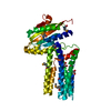
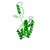
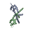
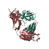
- PDB-1v7l: Structure of 3-isopropylmalate isomerase small subunit from Pyroc... -
+ Open data

- Basic information
| Entry | Database: PDB / ID: 1v7l | ||||||
|---|---|---|---|---|---|---|---|
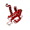
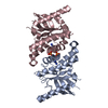
| Title | Structure of 3-isopropylmalate isomerase small subunit from Pyrococcus horikoshii | ||||||
 Components Components | 3-isopropylmalate dehydratase small subunit | ||||||
 Keywords Keywords | LYASE / BETA BARREL | ||||||
| Function / homology |  Function and homology information Function and homology information3-isopropylmalate dehydratase / 3-isopropylmalate dehydratase activity / L-leucine biosynthetic process Similarity search - Function | ||||||
| Biological species |   Pyrococcus horikoshii (archaea) Pyrococcus horikoshii (archaea) | ||||||
| Method |  X-RAY DIFFRACTION / SYNCHROTRON / MAD / Resolution: 1.98 Å X-RAY DIFFRACTION / SYNCHROTRON / MAD / Resolution: 1.98 Å | ||||||
 Authors Authors | Yao, M. / Kirita, T. / Sakai, N. / Tanaka, I. | ||||||
 Citation Citation | Journal: J.Mol.Biol. / Year: 2004 Title: Crystal Structure of the Pyrococcus horikoshii Isopropylmalate Isomerase Small Subunit Provides Insight into the Dual Substrate Specificity of the Enzyme Authors: Yasutake, Y. / Yao, M. / Sakai, N. / Kirita, T. / Tanaka, I. | ||||||
| History |
|
- Structure visualization
| Structure viewer | Molecule: MolmilJmol/JSmol |
|---|
- Downloads & links
-Download
| PDBx/mmCIF format | 1v7l.cif.gz | 80.4 KB | Display | PDBx/mmCIF format |
|---|---|---|---|---|
| PDB format | pdb1v7l.ent.gz | 61.6 KB | Display | PDB format |
| PDBx/mmJSON format | 1v7l.json.gz | Tree view | PDBx/mmJSON format | |
| Others |  Other downloads Other downloads |
-Validation report
| Summary document | 1v7l_validation.pdf.gz | 434.4 KB | Display | wwPDB validaton report |
|---|---|---|---|---|
| Full document | 1v7l_full_validation.pdf.gz | 445.3 KB | Display | |
| Data in XML | 1v7l_validation.xml.gz | 18.5 KB | Display | |
| Data in CIF | 1v7l_validation.cif.gz | 26.7 KB | Display | |
| Arichive directory | https://data.pdbj.org/pub/pdb/validation_reports/v7/1v7lftp://data.pdbj.org/pub/pdb/validation_reports/v7/1v7l | HTTPS FTP |
-Related structure data
| Similar structure data |
|---|

-Links
 PDBj
PDBj

- Assembly
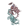
| Deposited unit | 
| ||||||||
|---|---|---|---|---|---|---|---|---|---|
| 1 |
| ||||||||
| 2 | 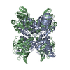
| ||||||||
| 3 | 
| ||||||||
| 4 | 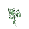
| ||||||||
| Unit cell |
|
-Components
| #1: Protein | Mass: 18033.779 Da / Num. of mol.: 2 Source method: isolated from a genetically manipulated source Source: (gene. exp.) Pyrococcus horikoshii (archaea) / Strain: OT3 / Gene: PH1724 / Plasmid: pET-22b / Production host:  #2: Water | ChemComp-HOH / |  Mass: 18.015 Da / Num. of mol.: 294 / Source method: isolated from a natural source / Formula: H2O Mass: 18.015 Da / Num. of mol.: 294 / Source method: isolated from a natural source / Formula: H2O |
|---|
-Experimental details
-Experiment
| Experiment | Method: X-RAY DIFFRACTION / Number of used crystals: 1 |
|---|
- Sample preparation
| Crystal | Density Matthews: 2.52 Å3/Da / Density % sol: 50.83 % |
|---|---|
| Crystal grow | Temperature: 293 K / Method: vapor diffusion, hanging drop / pH: 7.3 Details: 0.1M HEPES, 1.4M AMMONIUM SULFATE, pH 7.3, VAPOR DIFFUSION, HANGING DROP, temperature 293K |
-Data collection
| Diffraction | Mean temperature: 100 K | |||||||||||||||
|---|---|---|---|---|---|---|---|---|---|---|---|---|---|---|---|---|
| Diffraction source | Source: SYNCHROTRON / Site: SPring-8  / Beamline: BL41XU / Wavelength: 1.045 / Wavelength: 0.979131, 0.979359, 0.97001 / Beamline: BL41XU / Wavelength: 1.045 / Wavelength: 0.979131, 0.979359, 0.97001 | |||||||||||||||
| Detector | Type: MARRESEARCH / Detector: CCD / Date: Dec 19, 2000 | |||||||||||||||
| Radiation | Protocol: MAD / Monochromatic (M) / Laue (L): M / Scattering type: x-ray | |||||||||||||||
| Radiation wavelength |
| |||||||||||||||
| Reflection | Resolution: 1.98→25 Å / Num. all: 38097 / Num. obs: 29033 / % possible obs: 99.8 % / Observed criterion σ(I): 3 / Redundancy: 15.1 % / Biso Wilson estimate: 35.8 Å2 / Rmerge(I) obs: 0.084 / Rsym value: 0.082 / Net I/σ(I): 7.1 | |||||||||||||||
| Reflection shell | Resolution: 1.98→2.09 Å / Redundancy: 15.4 % / Rmerge(I) obs: 0.389 / Mean I/σ(I) obs: 2 / Num. unique all: 4159 / Rsym value: 0.377 / % possible all: 99.8 |
- Processing
| Software |
| |||||||||||||||||||||||||||
|---|---|---|---|---|---|---|---|---|---|---|---|---|---|---|---|---|---|---|---|---|---|---|---|---|---|---|---|---|
| Refinement | Method to determine structure: MAD / Resolution: 1.98→10 Å / Data cutoff low absF: 0 / Isotropic thermal model: Isotropic / Cross valid method: THROUGHOUT / σ(F): 0 / Stereochemistry target values: Engh & Huber
| |||||||||||||||||||||||||||
| Solvent computation | Solvent model: throughout / Bsol: 55.98 Å2 / ksol: 0.439 e/Å3 | |||||||||||||||||||||||||||
| Displacement parameters | Biso mean: 31.34 Å2
| |||||||||||||||||||||||||||
| Refine analyze |
| |||||||||||||||||||||||||||
| Refinement step | Cycle: LAST / Resolution: 1.98→10 Å
| |||||||||||||||||||||||||||
| Refine LS restraints |
| |||||||||||||||||||||||||||
| LS refinement shell | Resolution: 1.98→2.1 Å / Total num. of bins used: 10
| |||||||||||||||||||||||||||
| Xplor file | Serial no: 1 / Param file: CNS_PROTEIN_REP.PARAM / Topol file: CNS_PROTEIN.TOP |
