 Movie
Movie Controller


+ Open data

- Basic information
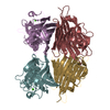
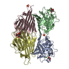
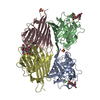
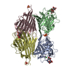
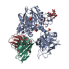
| Entry | Database: PDB / ID: 1v6n | ||||||
|---|---|---|---|---|---|---|---|
| Title | Peanut lectin with 9mer peptide (PVIWSSATG) | ||||||
 Components Components | Galactose-binding lectin | ||||||
 Keywords Keywords | SUGAR BINDING PROTEIN /  Lectin / agglutinin / open quaternary association / peptide and monoclinic Lectin / agglutinin / open quaternary association / peptide and monoclinic | ||||||
| Function / homology |  Function and homology information Function and homology information | ||||||
| Biological species |  Arachis hypogaea (peanut) Arachis hypogaea (peanut) | ||||||
| Method | X-RAY DIFFRACTION / Isomorphous replacement / Resolution: 3.5 Å | ||||||
 Authors Authors | Kundhavai Natchiar, S. / Arockia Jeyaprakash, A. / Ramya, T.N.C. / Thomas, C.J. / Suguna, K. / Surolia, A. / Vijayan, M. | ||||||
 Citation Citation | Journal: Acta Crystallogr.,Sect.D / Year: 2004 Title: Structural plasticity of peanut lectin: an X-ray analysis involving variation in pH, ligand binding and crystal structure. Authors: Kundhavai Natchiar, S. / Arockia Jeyaprakash, A. / Ramya, T.N. / Thomas, C.J. / Suguna, K. / Surolia, A. / Vijayan, M. #1: Journal: PROTEINS: STRUCT.,FUNCT.,GENET. / Year: 2001Title: Crystal structures of the peanut lectin-lactose complex at acidic pH: retention of unusual quaternary structure, empty and carbohydrate bound combining sites, molecular mimicry and crystal ...Title: Crystal structures of the peanut lectin-lactose complex at acidic pH: retention of unusual quaternary structure, empty and carbohydrate bound combining sites, molecular mimicry and crystal packing directed by interactions at the combining site Authors: Ravishankar, R. / Thomas, C.J. / Suguna, K. / Surolia, A. / Vijayan, M. | ||||||
| History |
|
- Structure visualization
| Structure viewer | Molecule: MolmilJmol/JSmol |
|---|
- Downloads & links
-Download
| PDBx/mmCIF format | 1v6n.cif.gz | 316.5 KB | Display | PDBx/mmCIF format |
|---|---|---|---|---|
| PDB format | pdb1v6n.ent.gz | 256.1 KB | Display | PDB format |
| PDBx/mmJSON format | 1v6n.json.gz | Tree view | PDBx/mmJSON format | |
| Others |  Other downloads Other downloads |
-Validation report
| Arichive directory | https://data.pdbj.org/pub/pdb/validation_reports/v6/1v6nftp://data.pdbj.org/pub/pdb/validation_reports/v6/1v6n | HTTPS FTP |
|---|
-Related structure data
| Related structure data |  1v6iC  1v6jC  1v6kC 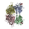 1v6lC  1v6mC  1v6oC  1cr7S S: Starting model for refinement C: citing same article ( |
|---|---|
| Similar structure data |


-Links
 PDBj
PDBj

- Assembly
| Deposited unit | 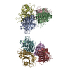
| ||||||||
|---|---|---|---|---|---|---|---|---|---|
| 1 | 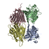
| ||||||||
| 2 | 
| ||||||||
| Unit cell |
|
-Components
| #1: Protein | Mass: 24706.385 Da / Num. of mol.: 8 / Source method: isolated from a natural source / Source: (natural) Arachis hypogaea (peanut) / Tissue: seed / References: UniProt: P02872#2: Chemical | ChemComp-CA /   Mass: 40.078 Da / Num. of mol.: 8 / Source method: obtained synthetically / Formula: Ca Mass: 40.078 Da / Num. of mol.: 8 / Source method: obtained synthetically / Formula: Ca#3: Chemical | ChemComp-MN /   Mass: 54.938 Da / Num. of mol.: 8 / Source method: obtained synthetically / Formula: Mn Mass: 54.938 Da / Num. of mol.: 8 / Source method: obtained synthetically / Formula: Mn#4: Water | ChemComp-HOH / | Water Mass: 18.015 Da / Num. of mol.: 207 / Source method: isolated from a natural source / Formula: H2O Mass: 18.015 Da / Num. of mol.: 207 / Source method: isolated from a natural source / Formula: H2O |
|---|
-Experimental details
-Experiment
| Experiment | Method: X-RAY DIFFRACTION / Number of used crystals: 1 |
|---|
- Sample preparation
| Crystal | Density Matthews: 3.11 Å3/Da / Density % sol: 60.14 % | ||||||||||||||||||||||||||||||||||||||||||
|---|---|---|---|---|---|---|---|---|---|---|---|---|---|---|---|---|---|---|---|---|---|---|---|---|---|---|---|---|---|---|---|---|---|---|---|---|---|---|---|---|---|---|---|
| Crystal grow | Temperature: 293 K / Method: hanging drop method / pH: 4.6 Details: 30% PEG8000, 0.2M Ammonium sulfate and 0.1M cacodylate, pH 4.6, hanging drop method, temperature 293K | ||||||||||||||||||||||||||||||||||||||||||
| Crystal grow | *PLUS Method: vapor diffusion, hanging drop | ||||||||||||||||||||||||||||||||||||||||||
| Components of the solutions | *PLUS
|
-Data collection
| Diffraction | Mean temperature: 293 K |
|---|---|
| Diffraction source | Source: ROTATING ANODE / Type: RIGAKU RU200 / Wavelength: 1.5418 Å |
| Detector | Type: MARRESEARCH / Detector: IMAGE PLATE / Details: Mirror |
| Radiation | Protocol: SINGLE WAVELENGTH / Monochromatic (M) / Laue (L): M / Scattering type: x-ray |
| Radiation wavelength | Wavelength: 1.5418 Å / Relative weight: 1 |
| Reflection | Resolution: 3.5→20 Å / Num. all: 30347 / Num. obs: 26281 / % possible obs: 94.7 % / Redundancy: 2.2 % / Rmerge(I) obs: 0.182 |
| Reflection shell | Resolution: 3.5→3.59 Å / Redundancy: 2.5 % / Rmerge(I) obs: 0.303 / Num. unique all: 775 / % possible all: 97.4 |
| Reflection | *PLUS Highest resolution: 3.5 Å / Num. measured all: 58800 |
| Reflection shell | *PLUS Highest resolution: 3.5 Å / % possible obs: 97.4 % / Num. unique obs: 775 / Num. measured obs: 1908 |
- Processing
| Software |
| ||||||||||||||||||||||||||||||||||||
|---|---|---|---|---|---|---|---|---|---|---|---|---|---|---|---|---|---|---|---|---|---|---|---|---|---|---|---|---|---|---|---|---|---|---|---|---|---|
| Refinement | Method to determine structure: Isomorphous replacement Starting model: 1CR7 Resolution: 3.5→20 Å / Rfactor Rfree error: 0.007 / Data cutoff high absF: 97064.81 / Data cutoff low absF: 0 / Cross valid method: THROUGHOUT / σ(F): 0
| ||||||||||||||||||||||||||||||||||||
| Solvent computation | Solvent model: FLAT MODEL / Bsol: 10 Å2 / ksol: 0.289869 e/Å3 | ||||||||||||||||||||||||||||||||||||
| Displacement parameters | Biso mean: 9.3 Å2
| ||||||||||||||||||||||||||||||||||||
| Refine analyze |
| ||||||||||||||||||||||||||||||||||||
| Refinement step | Cycle: LAST / Resolution: 3.5→20 Å
| ||||||||||||||||||||||||||||||||||||
| Refine LS restraints |
| ||||||||||||||||||||||||||||||||||||
| LS refinement shell | Resolution: 3.5→3.72 Å / Rfactor Rfree error: 0.021 / Total num. of bins used: 6
| ||||||||||||||||||||||||||||||||||||
| Xplor file |
| ||||||||||||||||||||||||||||||||||||
| Refinement | *PLUS Lowest resolution: 20 Å | ||||||||||||||||||||||||||||||||||||
| Solvent computation | *PLUS | ||||||||||||||||||||||||||||||||||||
| Displacement parameters | *PLUS | ||||||||||||||||||||||||||||||||||||
| Refine LS restraints | *PLUS
|
