 Movie
Movie Controller


+ Open data

- Basic information
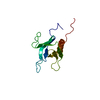
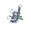
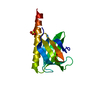
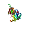
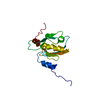
| Entry | Database: PDB / ID: 1v6b | ||||||
|---|---|---|---|---|---|---|---|
| Title | Solution structure of the third PDZ domain of mouse harmonin | ||||||
 Components Components | harmonin isoform a1 | ||||||
 Keywords Keywords |  PROTEIN BINDING / structural genomics / Usher syndrome / USH1 / RIKEN Structural Genomics/Proteomics Initiative / RSGI PROTEIN BINDING / structural genomics / Usher syndrome / USH1 / RIKEN Structural Genomics/Proteomics Initiative / RSGI | ||||||
| Function / homology |  Function and homology information Function and homology informationmyosin tail binding / protein-containing complex assembly => GO:0065003 / protein localization to microvillus / stereocilia ankle link / brush border assembly / regulation of microvillus length / stereocilia ankle link complex / parallel actin filament bundle assembly / retinal cone cell development / equilibrioception ...myosin tail binding / protein-containing complex assembly => GO:0065003 / protein localization to microvillus / stereocilia ankle link / brush border assembly / regulation of microvillus length / stereocilia ankle link complex / parallel actin filament bundle assembly / retinal cone cell development / equilibrioception / sensory perception of light stimulus / stereocilium tip / inner ear receptor cell stereocilium organization / inner ear auditory receptor cell differentiation / stereocilium / photoreceptor cell maintenance / inner ear morphogenesis / spectrin binding / microvillus / actin filament bundle assembly / brush border / neuromuscular process controlling balance / photoreceptor outer segment / photoreceptor inner segment / sensory perception of sound / G2/M transition of mitotic cell cycle / actin filament binding / apical part of cell / cytoskeleton / synapse / plasma membrane / cytosol / cytoplasmSimilarity search - Function | ||||||
| Biological species |  Mus musculus (house mouse) Mus musculus (house mouse) | ||||||
| Method | SOLUTION NMR | ||||||
 Authors Authors | Yamada, K. / Nameki, N. / Saito, K. / Koshiba, S. / Inoue, M. / Kigawa, T. / Yokoyama, S. / RIKEN Structural Genomics/Proteomics Initiative (RSGI) | ||||||
 Citation Citation | Journal: To be Published Title: Solution structure of the third PDZ domain of mouse harmonin Authors: Yamada, K. / Nameki, N. / Saito, K. / Koshiba, S. / Inoue, M. / Kigawa, T. / Yokoyama, S. | ||||||
| History |
|
- Structure visualization
| Structure viewer | Molecule: MolmilJmol/JSmol |
|---|
- Downloads & links
-Download
| PDBx/mmCIF format | 1v6b.cif.gz | 677.3 KB | Display | PDBx/mmCIF format |
|---|---|---|---|---|
| PDB format | pdb1v6b.ent.gz | 564.6 KB | Display | PDB format |
| PDBx/mmJSON format | 1v6b.json.gz | Tree view | PDBx/mmJSON format | |
| Others |  Other downloads Other downloads |
-Validation report
| Arichive directory | https://data.pdbj.org/pub/pdb/validation_reports/v6/1v6bftp://data.pdbj.org/pub/pdb/validation_reports/v6/1v6b | HTTPS FTP |
|---|
-Related structure data
| Similar structure data | |
|---|---|
| Other databases |


-Links
 PDBj
PDBj- Assembly
| Deposited unit | 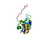
| |||||||||
|---|---|---|---|---|---|---|---|---|---|---|
| 1 |
| |||||||||
| NMR ensembles |
|
-Components
| #1: Protein | Mass: 12464.007 Da / Num. of mol.: 1 / Fragment: PDZ domain Source method: isolated from a genetically manipulated source Source: (gene. exp.) Mus musculus (house mouse) / Description: Cell-free protein synthesis / Gene: RIKEN cDNA 2010016F01 / Plasmid: P020401-47 / References: UniProt: Q9ES64 |
|---|
-Experimental details
-Experiment
| Experiment | Method: SOLUTION NMR | ||||||||||||
|---|---|---|---|---|---|---|---|---|---|---|---|---|---|
| NMR experiment |
|
- Sample preparation
| Details | Contents: 20mM Sodium Phosphate buffer, 100mM NaCl, 1mM d-DTT, 0.02% NaN3, 90% H2O, 10% D2O Solvent system: 90% H2O/10% D2O |
|---|---|
| Sample conditions | Ionic strength: 120mM / pH: 6.0 / Pressure: ambient / Temperature: 298 K |
-NMR measurement
| Radiation | Protocol: SINGLE WAVELENGTH / Monochromatic (M) / Laue (L): M |
|---|---|
| Radiation wavelength | Relative weight: 1 |
| NMR spectrometer | Type: Bruker AVANCE / Manufacturer: Bruker / Model: AVANCE / Field strength: 800 MHz |
- Processing
| NMR software |
| ||||||||||||||||||||||||||||
|---|---|---|---|---|---|---|---|---|---|---|---|---|---|---|---|---|---|---|---|---|---|---|---|---|---|---|---|---|---|
| NMR representative | Selection criteria: lowest energy | ||||||||||||||||||||||||||||
| NMR ensemble | Conformer selection criteria: target function / Conformers calculated total number: 100 / Conformers submitted total number: 20 |
