 Movie
Movie Controller

[English] 日本語
 Yorodumi
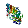
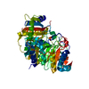
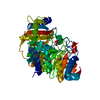
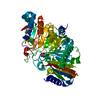
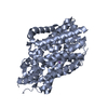
- PDB-1v0y: Phospholipase D from Streptomyces sp. strain PMF soaked with the ... -
+ Open data

- Basic information
| Entry | Database: PDB / ID: 1v0y | ||||||
|---|---|---|---|---|---|---|---|
| Title | Phospholipase D from Streptomyces sp. strain PMF soaked with the substrate dibutyrylphosphatidylcholine. | ||||||
 Components Components | PHOSPHOLIPASE D | ||||||
 Keywords Keywords | HYDROLASE / PHOSPHOLIPASE D / SUBSTRATE SOAK / DIBUTYRYLPHOSPHATIDYLCHOLINE / DIC4PC | ||||||
| Function / homology |  Function and homology information Function and homology informationphosphatidyltransferase activity / cardiolipin biosynthetic process / N-acylphosphatidylethanolamine-specific phospholipase D activity / phospholipase D / phospholipase D activitySimilarity search - Function | ||||||
| Biological species |  STREPTOMYCES SP. (bacteria) STREPTOMYCES SP. (bacteria) | ||||||
| Method | X-RAY DIFFRACTION / SYNCHROTRON / MOLECULAR REPLACEMENT / Resolution: 1.71 Å | ||||||
 Authors Authors | Leiros, I. / McSweeney, S. / Hough, E. | ||||||
 Citation Citation | Journal: J.Mol.Biol. / Year: 2004 Title: The Reaction Mechanism of Phospholipase D from Streptomyces Sp. Strain Pmf. Snapshots Along the Reaction Pathway Reveal a Pentacoordinate Reaction Intermediate and an Unexpected Final Product Authors: Leiros, I. / Mcsweeney, S. / Hough, E. #1: Journal: Structure / Year: 2000Title: The First Crystal Structure of a Phospholipase D Authors: Leiros, I. / Secundo, F. / Zambonelli, C. / Servi, S. / Hough, E. | ||||||
| History |
| ||||||
| Remark 700 | SHEET THE SHEET STRUCTURE OF THIS MOLECULE IS BIFURCATED. IN ORDER TO REPRESENT THIS FEATURE IN ... SHEET THE SHEET STRUCTURE OF THIS MOLECULE IS BIFURCATED. IN ORDER TO REPRESENT THIS FEATURE IN THE SHEET RECORDS BELOW, TWO SHEETS ARE DEFINED. |
- Structure visualization
| Structure viewer | Molecule: MolmilJmol/JSmol |
|---|
- Downloads & links
-Download
| PDBx/mmCIF format | 1v0y.cif.gz | 117.3 KB | Display | PDBx/mmCIF format |
|---|---|---|---|---|
| PDB format | pdb1v0y.ent.gz | 88 KB | Display | PDB format |
| PDBx/mmJSON format | 1v0y.json.gz | Tree view | PDBx/mmJSON format | |
| Others |  Other downloads Other downloads |
-Validation report
| Arichive directory | https://data.pdbj.org/pub/pdb/validation_reports/v0/1v0yftp://data.pdbj.org/pub/pdb/validation_reports/v0/1v0y | HTTPS FTP |
|---|
-Related structure data
| Related structure data |  1v0rC  1v0sC 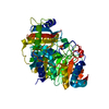 1v0tC  1v0uC  1v0vC  1v0wC  1f0iS C: citing same article ( S: Starting model for refinement |
|---|---|
| Similar structure data |


-Links
 PDBj
PDBj- Assembly
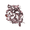
| Deposited unit | 
| ||||||||
|---|---|---|---|---|---|---|---|---|---|
| 1 |
| ||||||||
| Unit cell |
|
-Components
| #1: Protein | Mass: 54064.453 Da / Num. of mol.: 1 / Source method: isolated from a natural source / Source: (natural) STREPTOMYCES SP. (bacteria) / Strain: PMF / References: UniProt: P84147*PLUS, phospholipase D |
|---|---|
| #2: Chemical | ChemComp-HI5 / 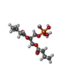  Mass: 330.269 Da / Num. of mol.: 1 / Source method: obtained synthetically / Formula: C11H23O9P Mass: 330.269 Da / Num. of mol.: 1 / Source method: obtained synthetically / Formula: C11H23O9P |
| #3: Water | ChemComp-HOH / Water Mass: 18.015 Da / Num. of mol.: 413 / Source method: isolated from a natural source / Formula: H2O Mass: 18.015 Da / Num. of mol.: 413 / Source method: isolated from a natural source / Formula: H2O |
| Compound details | THIS DATASET DESCRIBES A 30 MINUTE SUBSTRATE SOAK USING 2MM DIBUTYRYLPHOSPHATIDYLCHOLINE. ...THIS DATASET DESCRIBES A 30 MINUTE SUBSTRATE SOAK USING 2MM DIBUTYRYLP |
| Sequence details | THIS SEQUENCE HAS NOT BEEN DEPOSITED TO UNIPROT AT THE TIME OF STRUCTURE DEPOSITION |
-Experimental details
-Experiment
| Experiment | Method: X-RAY DIFFRACTION / Number of used crystals: 1 |
|---|
- Sample preparation
| Crystal | Density Matthews: 2 Å3/Da / Density % sol: 38.4 % |
|---|---|
| Crystal grow | pH: 5.4 Details: 0.2 M NH4AC, 0.1 M CITRATE PHOSPHATE BUFFER AT PH5.4, 27.5% PEG 4000. CRYSTALS WERE THEREAFTER BACKSOAKED TWICE IN A PHOSPHATE- FREE BUFFER TO REMOVE TRACE-AMOUNTS OF PHOSPHATE., pH 5.40 |
-Data collection
| Diffraction | Mean temperature: 100 K |
|---|---|
| Diffraction source | Source: SYNCHROTRON / Site: ESRF  / Beamline: BM1A / Wavelength: 0.873 / Beamline: BM1A / Wavelength: 0.873 |
| Detector | Type: MARRESEARCH / Detector: IMAGE PLATE |
| Radiation | Protocol: SINGLE WAVELENGTH / Monochromatic (M) / Laue (L): M / Scattering type: x-ray |
| Radiation wavelength | Wavelength: 0.873 Å / Relative weight: 1 |
| Reflection | Resolution: 1.7→13.71 Å / Num. obs: 45138 / % possible obs: 99.2 % / Observed criterion σ(I): 0 / Redundancy: 4.9 % / Rmerge(I) obs: 0.115 / Net I/σ(I): 4.5 |
| Reflection shell | Resolution: 1.7→1.8 Å / Redundancy: 4.6 % / Rmerge(I) obs: 0.473 / Mean I/σ(I) obs: 2 / % possible all: 92.9 |
- Processing
| Software |
| ||||||||||||||||||||||||||||||||||||||||||||||||||||||||||||||||||||||||||||||||||||||||||||||||||||||||||||||||||||||||||||||||||||||||||||||||||||||||||||||||||||||||||||||||||||||
|---|---|---|---|---|---|---|---|---|---|---|---|---|---|---|---|---|---|---|---|---|---|---|---|---|---|---|---|---|---|---|---|---|---|---|---|---|---|---|---|---|---|---|---|---|---|---|---|---|---|---|---|---|---|---|---|---|---|---|---|---|---|---|---|---|---|---|---|---|---|---|---|---|---|---|---|---|---|---|---|---|---|---|---|---|---|---|---|---|---|---|---|---|---|---|---|---|---|---|---|---|---|---|---|---|---|---|---|---|---|---|---|---|---|---|---|---|---|---|---|---|---|---|---|---|---|---|---|---|---|---|---|---|---|---|---|---|---|---|---|---|---|---|---|---|---|---|---|---|---|---|---|---|---|---|---|---|---|---|---|---|---|---|---|---|---|---|---|---|---|---|---|---|---|---|---|---|---|---|---|---|---|---|---|
| Refinement | Method to determine structure: MOLECULAR REPLACEMENT Starting model: BASED ON PDB ENTRY 1F0I Resolution: 1.71→13.71 Å / Cor.coef. Fo:Fc: 0.966 / Cor.coef. Fo:Fc free: 0.953 / SU B: 2.298 / SU ML: 0.073 / Cross valid method: THROUGHOUT / ESU R: 0.104 / ESU R Free: 0.105 / Stereochemistry target values: MAXIMUM LIKELIHOOD Details: HYDROGENS HAVE BEEN ADDED IN THE RIDING POSITIONS. THIS ENTRY CONTAINS ATOMS THAT HAVE BEEN MODELED WITH AN OCCUPANCY OF 0.00.
| ||||||||||||||||||||||||||||||||||||||||||||||||||||||||||||||||||||||||||||||||||||||||||||||||||||||||||||||||||||||||||||||||||||||||||||||||||||||||||||||||||||||||||||||||||||||
| Solvent computation | Ion probe radii: 0.8 Å / Shrinkage radii: 0.8 Å / VDW probe radii: 1.4 Å / Solvent model: BABINET MODEL WITH MASK | ||||||||||||||||||||||||||||||||||||||||||||||||||||||||||||||||||||||||||||||||||||||||||||||||||||||||||||||||||||||||||||||||||||||||||||||||||||||||||||||||||||||||||||||||||||||
| Displacement parameters | Biso mean: 20.18 Å2
| ||||||||||||||||||||||||||||||||||||||||||||||||||||||||||||||||||||||||||||||||||||||||||||||||||||||||||||||||||||||||||||||||||||||||||||||||||||||||||||||||||||||||||||||||||||||
| Refinement step | Cycle: LAST / Resolution: 1.71→13.71 Å
| ||||||||||||||||||||||||||||||||||||||||||||||||||||||||||||||||||||||||||||||||||||||||||||||||||||||||||||||||||||||||||||||||||||||||||||||||||||||||||||||||||||||||||||||||||||||
| Refine LS restraints |
|