 Movie
Movie Controller


+ Open data

- Basic information
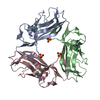
| Entry | Database: PDB / ID: 1usz | ||||||
|---|---|---|---|---|---|---|---|
| Title | SeMet AfaE-3 adhesin from Escherichia Coli | ||||||
 Components Components | AFIMBRIAL ADHESIN AFA-III | ||||||
 Keywords Keywords | ADHESIN / AFAE-3 / AFIMBRIAL ADHESIN / UPEC / DAEC | ||||||
| Function / homology |  Function and homology information Function and homology information | ||||||
| Biological species |   ESCHERICHIA COLI (E. coli) ESCHERICHIA COLI (E. coli) | ||||||
| Method | X-RAY DIFFRACTION / SYNCHROTRON / MAD / Resolution: 3.28 Å | ||||||
 Authors Authors | Anderson, K.L. / Billington, J. / Pettigrew, D. / Cota, E. / Roversi, P. / Simpson, P. / Chen, H.A. / Urvil, P. / Dumerle, L. / Barlow, P. ...Anderson, K.L. / Billington, J. / Pettigrew, D. / Cota, E. / Roversi, P. / Simpson, P. / Chen, H.A. / Urvil, P. / Dumerle, L. / Barlow, P. / Medof, E. / Smith, R.A.G. / Nowicki, B. / Le Bouguenec, C. / Lea, S.M. / Matthews, S. | ||||||
 Citation Citation | Journal: J.Biol.Chem. / Year: 2004 Title: High Resolution Studies of the Afa/Dr Adhesin Drae and its Interaction with Chloramphenicol Authors: Pettigrew, D. / Anderson, K.L. / Billington, J. / Cota, E. / Simpson, P. / Urvil, P. / Rabuzin, F. / Roversi, P. / Nowicki, B. / Du Merle, L. / Le Bouguenec, C. / Matthews, S. / Lea, S.M. #1: Journal: Mol.Cell / Year: 2004Title: An Atomic Resolution Model for Assmebly, Architecture,and Function of the Dr Adhesins Authors: Anderson, K.L. / Billington, J. / Pettigrew, D. / Cota, E. / Simpson, P. / Roversi, P. / Chen, H.A. / Urvil, P. / Du Merle, L. / Barlow, P.N. / Medof, M.E. / Smith, R.A.G. / Nowicki, B. / Le ...Authors: Anderson, K.L. / Billington, J. / Pettigrew, D. / Cota, E. / Simpson, P. / Roversi, P. / Chen, H.A. / Urvil, P. / Du Merle, L. / Barlow, P.N. / Medof, M.E. / Smith, R.A.G. / Nowicki, B. / Le Bouguenec, C. / Lea, S.M. / Matthews, S. | ||||||
| History |
| ||||||
| Remark 700 | SHEET THE SHEET STRUCTURE OF THIS MOLECULE IS BIFURCATED. IN ORDER TO REPRESENT THIS FEATURE IN ... SHEET THE SHEET STRUCTURE OF THIS MOLECULE IS BIFURCATED. IN ORDER TO REPRESENT THIS FEATURE IN THE SHEET RECORDS BELOW, TWO SHEETS ARE DEFINED. |
- Structure visualization
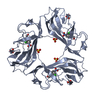
| Structure viewer | Molecule: MolmilJmol/JSmol |
|---|
- Downloads & links
-Download
| PDBx/mmCIF format | 1usz.cif.gz | 35.9 KB | Display | PDBx/mmCIF format |
|---|---|---|---|---|
| PDB format | pdb1usz.ent.gz | 28.3 KB | Display | PDB format |
| PDBx/mmJSON format | 1usz.json.gz | Tree view | PDBx/mmJSON format | |
| Others |  Other downloads Other downloads |
-Validation report
| Arichive directory | https://data.pdbj.org/pub/pdb/validation_reports/us/1uszftp://data.pdbj.org/pub/pdb/validation_reports/us/1usz | HTTPS FTP |
|---|
-Related structure data


-Links
 PDBj
PDBj- Assembly
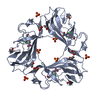
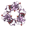
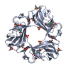
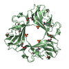
| Deposited unit | 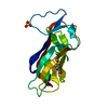
| ||||||||
|---|---|---|---|---|---|---|---|---|---|
| 1 | 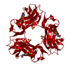
| ||||||||
| Unit cell |
| ||||||||
| Details | THE MONOMER SITS AROUND THE THREEFOLD AXIS AND FORMS ATRIMER WITH ITS THREEFOLD-RELATED COPIES; THE TRIMER ISLINKED INTERNALLY BY INTERMOLECULAR DISULPHIDE BONDS. |
-Components
| #1: Protein | Mass: 16233.725 Da / Num. of mol.: 1 / Mutation: YES Source method: isolated from a genetically manipulated source Details: SELENOMETHIONINE DERIVATIVE / Source: (gene. exp.) ESCHERICHIA COLI (E. coli) / Strain: A30 / Description: N-TERMINUS 6-HIS-TAGGED / Plasmid: PQE-30 / Production host: ESCHERICHIA COLI (E. coli) / Strain (production host): B834 / References: UniProt: Q57254 | ||
|---|---|---|---|
| #2: Chemical | ChemComp-SO4 / Sulfate  Mass: 96.063 Da / Num. of mol.: 1 / Source method: obtained synthetically / Formula: SO4 Mass: 96.063 Da / Num. of mol.: 1 / Source method: obtained synthetically / Formula: SO4 | ||
| #3: Chemical | ChemComp-CL / Chloride  Mass: 35.453 Da / Num. of mol.: 1 / Source method: obtained synthetically / Formula: Cl Mass: 35.453 Da / Num. of mol.: 1 / Source method: obtained synthetically / Formula: Cl | ||
| #4: Water | ChemComp-HOH / Water Mass: 18.015 Da / Num. of mol.: 4 / Source method: isolated from a natural source / Formula: H2O Mass: 18.015 Da / Num. of mol.: 4 / Source method: isolated from a natural source / Formula: H2O | ||
| Compound details | ENGINEERED| Sequence details | MUTATIONS A0G, G1S | |
-Experimental details
-Experiment
| Experiment | Method: X-RAY DIFFRACTION / Number of used crystals: 1 |
|---|
- Sample preparation
| Crystal | Density Matthews: 3.62 Å3/Da / Density % sol: 67 % / Description: SOLVED BY SAD WITH AUTOSHARP |
|---|---|
| Crystal grow | pH: 7 Details: 2.0 M AMMONIUM SULPHATE, 2% PEG 400, 0.1M NAHEPES PH 6.8 |
-Data collection
| Diffraction | Mean temperature: 100 K |
|---|---|
| Diffraction source | Source: SYNCHROTRON / Site: ESRF  / Beamline: BM14 / Wavelength: 0.97892 / Beamline: BM14 / Wavelength: 0.97892 |
| Detector | Type: MARRESEARCH / Detector: CCD / Date: Mar 15, 2003 |
| Radiation | Protocol: SINGLE WAVELENGTH / Monochromatic (M) / Laue (L): M / Scattering type: x-ray |
| Radiation wavelength | Wavelength: 0.97892 Å / Relative weight: 1 |
| Reflection | Resolution: 3.1→28.9 Å / Num. obs: 5108 / % possible obs: 99.7 % / Redundancy: 23 % / Biso Wilson estimate: -2.3 Å2 / Rmerge(I) obs: 0.084 / Net I/σ(I): 7.8 |
| Reflection shell | Resolution: 3.1→3.27 Å / Rmerge(I) obs: 0.32 / Mean I/σ(I) obs: 1.7 / % possible all: 100 |
- Processing
| Software |
| |||||||||||||||||||||||||||||||||||||||||||||||||||||||||||||||||||||||||||||||||||||||||||||||
|---|---|---|---|---|---|---|---|---|---|---|---|---|---|---|---|---|---|---|---|---|---|---|---|---|---|---|---|---|---|---|---|---|---|---|---|---|---|---|---|---|---|---|---|---|---|---|---|---|---|---|---|---|---|---|---|---|---|---|---|---|---|---|---|---|---|---|---|---|---|---|---|---|---|---|---|---|---|---|---|---|---|---|---|---|---|---|---|---|---|---|---|---|---|---|---|---|
| Refinement | Method to determine structure: MAD / Resolution: 3.28→30 Å / Isotropic thermal model: TNT BCORREL / Cross valid method: THROUGHOUT / σ(F): 0 / Stereochemistry target values: TNT CSDX_PROTGEO / Details: MAXIMUM LIKELIHOOD BUSTER-TNT REFINEMENT
| |||||||||||||||||||||||||||||||||||||||||||||||||||||||||||||||||||||||||||||||||||||||||||||||
| Solvent computation | Solvent model: BABINET SCALING / Bsol: 37 Å2 / ksol: 0.36 e/Å3 | |||||||||||||||||||||||||||||||||||||||||||||||||||||||||||||||||||||||||||||||||||||||||||||||
| Refinement step | Cycle: LAST / Resolution: 3.28→30 Å
| |||||||||||||||||||||||||||||||||||||||||||||||||||||||||||||||||||||||||||||||||||||||||||||||
| Refine LS restraints |
|
