 Movie
Movie Controller


+ Open data

- Basic information
| Entry | Database: PDB / ID: 1ur3 | ||||||
|---|---|---|---|---|---|---|---|
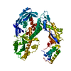
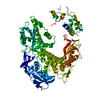
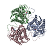
| Title | Crystal structure of the apo form of the E.coli ydhF protein | ||||||
 Components Components | HYPOTHETICAL OXIDOREDUCTASE YDHF | ||||||
 Keywords Keywords |  OXIDOREDUCTASE / NADP BINDING / ALDO-KETO REDUCTASE OXIDOREDUCTASE / NADP BINDING / ALDO-KETO REDUCTASE | ||||||
| Function / homology |  Function and homology information Function and homology information | ||||||
| Biological species |  ESCHERICHIA COLI (E. coli) ESCHERICHIA COLI (E. coli) | ||||||
| Method | X-RAY DIFFRACTION / SYNCHROTRON / MAD / Resolution: 2.57 Å | ||||||
 Authors Authors | Jeudy, S. / Claverie, J.M. / Abergel, C. | ||||||
 Citation Citation | Journal: To be Published Title: Crystal Structure of Ydhf the E.Coli Aldo-Keto Reductase Ydhf Authors: Jeudy, S. / Claverie, J.M. / Abergel, C. | ||||||
| History |
| ||||||
| Remark 700 | SHEET DETERMINATION METHOD: DSSP THE SHEETS PRESENTED AS "MB" IN EACH CHAIN ON SHEET RECORDS BELOW ... SHEET DETERMINATION METHOD: DSSP THE SHEETS PRESENTED AS "MB" IN EACH CHAIN ON SHEET RECORDS BELOW IS ACTUALLY AN 8-STRANDED BARREL THIS IS REPRESENTED BY A 9-STRANDED SHEET IN WHICH THE FIRST AND LAST STRANDS ARE IDENTICAL. |
- Structure visualization
| Structure viewer | Molecule: MolmilJmol/JSmol |
|---|
- Downloads & links
-Download
| PDBx/mmCIF format | 1ur3.cif.gz | 70.3 KB | Display | PDBx/mmCIF format |
|---|---|---|---|---|
| PDB format | pdb1ur3.ent.gz | 55.9 KB | Display | PDB format |
| PDBx/mmJSON format | 1ur3.json.gz | Tree view | PDBx/mmJSON format | |
| Others |  Other downloads Other downloads |
-Validation report
| Arichive directory | https://data.pdbj.org/pub/pdb/validation_reports/ur/1ur3ftp://data.pdbj.org/pub/pdb/validation_reports/ur/1ur3 | HTTPS FTP |
|---|
-Related structure data
| Related structure data | |
|---|---|
| Similar structure data |


-Links
 PDBj
PDBj
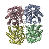
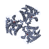
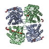
- Assembly
| Deposited unit | 
| ||||||||
|---|---|---|---|---|---|---|---|---|---|
| 1 | 
| ||||||||
| Unit cell |
|
-Components
| #1: Protein | Mass: 36577.512 Da / Num. of mol.: 1 Source method: isolated from a genetically manipulated source Source: (gene. exp.) ESCHERICHIA COLI (E. coli) / Strain: K12 / Production host: ESCHERICHIA COLI (E. coli) / Strain (production host): BL21(DE3) / References: UniProt: P76187, Oxidoreductases |
|---|---|
| #2: Chemical | ChemComp-SO4 / Sulfate  Mass: 96.063 Da / Num. of mol.: 1 / Source method: obtained synthetically / Formula: SO4 Mass: 96.063 Da / Num. of mol.: 1 / Source method: obtained synthetically / Formula: SO4 |
| #3: Water | ChemComp-HOH / Water Mass: 18.015 Da / Num. of mol.: 71 / Source method: isolated from a natural source / Formula: H2O Mass: 18.015 Da / Num. of mol.: 71 / Source method: isolated from a natural source / Formula: H2O |
| Compound details | BELONGS TO THE ALDO/KETO REDUCTASE 2 FAMILY. |
| Sequence details | FIRST METHIONINE WAS REPLACED WITH A LEUCINE ADDITION OF 21 N-TER RESIDUES CORRESPONDING TO THE ...FIRST METHIONINE |
-Experimental details
-Experiment
| Experiment | Method: X-RAY DIFFRACTION / Number of used crystals: 1 |
|---|
- Sample preparation
| Crystal | Density Matthews: 2.05 Å3/Da / Density % sol: 39.8 % |
|---|---|
| Crystal grow | pH: 7 Details: PEG 4000 17.5%, IMIDAZOLE/MALATE 0.2M PH 7.0, AMMONIUM SULFATE 0.1M, GLYCEROL 2.5%, SPERMIDINE 0.1M |
-Data collection
| Diffraction | Mean temperature: 105 K | ||||||||||||
|---|---|---|---|---|---|---|---|---|---|---|---|---|---|
| Diffraction source | Source: SYNCHROTRON / Site: ESRF  / Beamline: BM30A / Wavelength: 0.97917,0.98735,0.96 / Beamline: BM30A / Wavelength: 0.97917,0.98735,0.96 | ||||||||||||
| Detector | Type: MARRESEARCH / Detector: CCD / Date: Mar 15, 2002 / Details: MIRRORS | ||||||||||||
| Radiation | Protocol: MAD / Monochromatic (M) / Laue (L): M / Scattering type: x-ray | ||||||||||||
| Radiation wavelength |
| ||||||||||||
| Reflection | Resolution: 2.57→16 Å / Num. obs: 9276 / % possible obs: 98.7 % / Redundancy: 5.4 % / Biso Wilson estimate: 57.1 Å2 / Rmerge(I) obs: 0.078 / Net I/σ(I): 6.3 | ||||||||||||
| Reflection shell | Resolution: 2.57→2.66 Å / Redundancy: 2.9 % / Rmerge(I) obs: 0.322 / Mean I/σ(I) obs: 1.7 / % possible all: 98.7 |
- Processing
| Software |
| ||||||||||||||||||||||||||||||||||||||||||||||||||||||||||||||||||||||||||||||||
|---|---|---|---|---|---|---|---|---|---|---|---|---|---|---|---|---|---|---|---|---|---|---|---|---|---|---|---|---|---|---|---|---|---|---|---|---|---|---|---|---|---|---|---|---|---|---|---|---|---|---|---|---|---|---|---|---|---|---|---|---|---|---|---|---|---|---|---|---|---|---|---|---|---|---|---|---|---|---|---|---|---|
| Refinement | Method to determine structure: MAD / Resolution: 2.57→16.08 Å / Rfactor Rfree error: 0.008 / Data cutoff high absF: 2164594.45 / Isotropic thermal model: RESTRAINED / Cross valid method: THROUGHOUT / σ(F): 0
| ||||||||||||||||||||||||||||||||||||||||||||||||||||||||||||||||||||||||||||||||
| Solvent computation | Solvent model: FLAT MODEL / Bsol: 53.8829 Å2 / ksol: 0.368591 e/Å3 | ||||||||||||||||||||||||||||||||||||||||||||||||||||||||||||||||||||||||||||||||
| Displacement parameters | Biso mean: 38.6 Å2
| ||||||||||||||||||||||||||||||||||||||||||||||||||||||||||||||||||||||||||||||||
| Refine analyze |
| ||||||||||||||||||||||||||||||||||||||||||||||||||||||||||||||||||||||||||||||||
| Refinement step | Cycle: LAST / Resolution: 2.57→16.08 Å
| ||||||||||||||||||||||||||||||||||||||||||||||||||||||||||||||||||||||||||||||||
| Refine LS restraints |
| ||||||||||||||||||||||||||||||||||||||||||||||||||||||||||||||||||||||||||||||||
| LS refinement shell | Resolution: 2.57→2.73 Å / Rfactor Rfree error: 0.027 / Total num. of bins used: 6
| ||||||||||||||||||||||||||||||||||||||||||||||||||||||||||||||||||||||||||||||||
| Xplor file |
|
