 Movie
Movie Controller

[English] 日本語
 Yorodumi
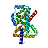
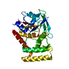
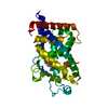
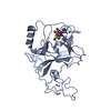
- PDB-1upv: Crystal structure of the human Liver X receptor beta ligand bindi... -
+ Open data

- Basic information
| Entry | Database: PDB / ID: 1upv | ||||||
|---|---|---|---|---|---|---|---|
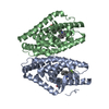
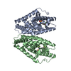
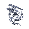
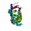
| Title | Crystal structure of the human Liver X receptor beta ligand binding domain in complex with a synthetic agonist | ||||||
 Components Components | OXYSTEROLS RECEPTOR LXR-BETA | ||||||
 Keywords Keywords |  RECEPTOR / NUCLEAR HORMONE RECEPTOR / LIGAND BINDING DOMAIN / LIVER X RECEPTOR / TRANSCRIPTION FACTOR RECEPTOR / NUCLEAR HORMONE RECEPTOR / LIGAND BINDING DOMAIN / LIVER X RECEPTOR / TRANSCRIPTION FACTOR | ||||||
| Function / homology |  Function and homology information Function and homology informationpositive regulation of secretion of lysosomal enzymes / positive regulation of high-density lipoprotein particle assembly / positive regulation of pancreatic juice secretion / phosphatidylcholine acyl-chain remodeling / positive regulation of cholesterol transport / negative regulation of response to endoplasmic reticulum stress / negative regulation of pinocytosis / positive regulation of lipoprotein lipase activity / apolipoprotein A-I receptor binding / positive regulation of triglyceride biosynthetic process ...positive regulation of secretion of lysosomal enzymes / positive regulation of high-density lipoprotein particle assembly / positive regulation of pancreatic juice secretion / phosphatidylcholine acyl-chain remodeling / positive regulation of cholesterol transport / negative regulation of response to endoplasmic reticulum stress / negative regulation of pinocytosis / positive regulation of lipoprotein lipase activity / apolipoprotein A-I receptor binding / positive regulation of triglyceride biosynthetic process / NR1H2 & NR1H3 regulate gene expression linked to triglyceride lipolysis in adipose / NR1H2 & NR1H3 regulate gene expression to limit cholesterol uptake / positive regulation of lipid storage / NR1H2 & NR1H3 regulate gene expression linked to lipogenesis / positive regulation of fatty acid biosynthetic process / negative regulation of lipid transport / negative regulation of cold-induced thermogenesis / NR1H2 & NR1H3 regulate gene expression to control bile acid homeostasis / negative regulation of type II interferon-mediated signaling pathway / negative regulation of cholesterol storage / negative regulation of macrophage derived foam cell differentiation / positive regulation of cholesterol efflux / nuclear retinoid X receptor binding / NR1H3 & NR1H2 regulate gene expression linked to cholesterol transport and efflux / positive regulation of protein metabolic process / VLDLR internalisation and degradation / hormone-mediated signaling pathway / cholesterol homeostasis / negative regulation of proteolysis / SUMOylation of intracellular receptors / mRNA transcription by RNA polymerase II / PPARA activates gene expression / chromatin DNA binding / positive regulation of miRNA transcription / Nuclear Receptor transcription pathway / RNA polymerase II transcription regulator complex / nuclear receptor activity / ATPase binding / DNA-binding transcription activator activity, RNA polymerase II-specific / cell differentiation / DNA-binding transcription factor activity, RNA polymerase II-specific / RNA polymerase II cis-regulatory region sequence-specific DNA binding / negative regulation of DNA-templated transcription / chromatin / positive regulation of DNA-templated transcription / negative regulation of transcription by RNA polymerase II / positive regulation of transcription by RNA polymerase II / DNA binding / zinc ion binding / nucleoplasm / nucleus / cytosol / cytoplasmSimilarity search - Function | ||||||
| Biological species |  HOMO SAPIENS (human) HOMO SAPIENS (human) | ||||||
| Method | X-RAY DIFFRACTION / SYNCHROTRON / MOLECULAR REPLACEMENT / Resolution: 2.1 Å | ||||||
 Authors Authors | Hoerer, S. / Schmid, A. / Heckel, A. / Budzinski, R.M. / Nar, H. | ||||||
 Citation Citation | Journal: J.Mol.Biol. / Year: 2003 Title: Crystal Structure of the Human Liver X Receptor Beta Ligand-Binding Domain in Complex with a Synthetic Agonist Authors: Hoerer, S. / Schmid, A. / Heckel, A. / Budzinski, R.M. / Nar, H. | ||||||
| History |
|
- Structure visualization
| Structure viewer | Molecule: MolmilJmol/JSmol |
|---|
- Downloads & links
-Download
| PDBx/mmCIF format | 1upv.cif.gz | 63.6 KB | Display | PDBx/mmCIF format |
|---|---|---|---|---|
| PDB format | pdb1upv.ent.gz | 45.3 KB | Display | PDB format |
| PDBx/mmJSON format | 1upv.json.gz | Tree view | PDBx/mmJSON format | |
| Others |  Other downloads Other downloads |
-Validation report
| Arichive directory | https://data.pdbj.org/pub/pdb/validation_reports/up/1upvftp://data.pdbj.org/pub/pdb/validation_reports/up/1upv | HTTPS FTP |
|---|
-Related structure data
| Related structure data |  1upwC  1fcyS C: citing same article ( S: Starting model for refinement |
|---|---|
| Similar structure data |


-Links
 PDBj
PDBj


- Assembly
| Deposited unit | 
| ||||||||
|---|---|---|---|---|---|---|---|---|---|
| 1 |
| ||||||||
| Unit cell |
|
-Components
| #1: Protein | Mass: 29416.621 Da / Num. of mol.: 1 / Fragment: LIGAND BINDING DOMAIN, RESIDUES 209-461 Source method: isolated from a genetically manipulated source Source: (gene. exp.) HOMO SAPIENS (human) / Plasmid: PET28A(+) / Production host:  Escherichia coli BL21(DE3) (bacteria) / References: UniProt: P55055 Escherichia coli BL21(DE3) (bacteria) / References: UniProt: P55055 |
|---|---|
| #2: Chemical | ChemComp-444 / 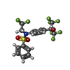  Mass: 481.333 Da / Num. of mol.: 1 / Source method: obtained synthetically / Formula: C17H12F9NO3S Mass: 481.333 Da / Num. of mol.: 1 / Source method: obtained synthetically / Formula: C17H12F9NO3S |
| #3: Water | ChemComp-HOH / Water Mass: 18.015 Da / Num. of mol.: 97 / Source method: isolated from a natural source / Formula: H2O Mass: 18.015 Da / Num. of mol.: 97 / Source method: isolated from a natural source / Formula: H2O |
| Compound details | FUNCTION: ORPHAN RECEPTOR. BINDS PREFERENTIALLY TO DOUBLE-STRANDED OLIGONUCLEOTIDE DIRECT REPEATS ...FUNCTION: ORPHAN RECEPTOR. BINDS PREFERENTI |
-Experimental details
-Experiment
| Experiment | Method: X-RAY DIFFRACTION / Number of used crystals: 1 |
|---|
- Sample preparation
| Crystal | Density Matthews: 2.6 Å3/Da / Density % sol: 52.3 % |
|---|---|
| Crystal grow | pH: 7.5 Details: RESERVOIR: 25% MPD, 10% PEG4K, 100 MM HEPES PH 7.5, 10 MM D PROTEIN: 4-8 MG/ML IN 20 MM TRIS PH 8.0, 100 MM NACL, 10 MM |
-Data collection
| Diffraction | Mean temperature: 100 K |
|---|---|
| Diffraction source | Source: SYNCHROTRON / Site: SLS  / Beamline: X06SA / Wavelength: 0.9166 / Beamline: X06SA / Wavelength: 0.9166 |
| Detector | Type: MARRESEARCH / Detector: CCD / Date: Oct 15, 2002 / Details: MIRRORS |
| Radiation | Monochromator: SI(111) MONOCHROMATOR / Protocol: SINGLE WAVELENGTH / Monochromatic (M) / Laue (L): M / Scattering type: x-ray |
| Radiation wavelength | Wavelength: 0.9166 Å / Relative weight: 1 |
| Reflection | Resolution: 2.1→30 Å / Num. obs: 15111 / % possible obs: 83.3 % / Redundancy: 2.6 % / Rmerge(I) obs: 0.093 / Net I/σ(I): 6.4 |
| Reflection shell | Resolution: 2.1→2.18 Å / Redundancy: 2.2 % / Rmerge(I) obs: 0.317 / Mean I/σ(I) obs: 2 / % possible all: 71.3 |
- Processing
| Software |
| ||||||||||||||||||||||||||||||||||||||||||||||||||||||||||||||||||||||||||||||||||||||||||||||||||||||||||||||||||||||||||||||||||||||||||||||||||||||||||||||||||||||||||||||||||||||
|---|---|---|---|---|---|---|---|---|---|---|---|---|---|---|---|---|---|---|---|---|---|---|---|---|---|---|---|---|---|---|---|---|---|---|---|---|---|---|---|---|---|---|---|---|---|---|---|---|---|---|---|---|---|---|---|---|---|---|---|---|---|---|---|---|---|---|---|---|---|---|---|---|---|---|---|---|---|---|---|---|---|---|---|---|---|---|---|---|---|---|---|---|---|---|---|---|---|---|---|---|---|---|---|---|---|---|---|---|---|---|---|---|---|---|---|---|---|---|---|---|---|---|---|---|---|---|---|---|---|---|---|---|---|---|---|---|---|---|---|---|---|---|---|---|---|---|---|---|---|---|---|---|---|---|---|---|---|---|---|---|---|---|---|---|---|---|---|---|---|---|---|---|---|---|---|---|---|---|---|---|---|---|---|
| Refinement | Method to determine structure: MOLECULAR REPLACEMENT Starting model: PDB ENTRY 1FCY Resolution: 2.1→20 Å / Cor.coef. Fo:Fc: 0.927 / Cor.coef. Fo:Fc free: 0.896 / SU B: 6.541 / SU ML: 0.171 / Cross valid method: THROUGHOUT / ESU R: 0.278 / ESU R Free: 0.234 / Stereochemistry target values: MAXIMUM LIKELIHOOD
| ||||||||||||||||||||||||||||||||||||||||||||||||||||||||||||||||||||||||||||||||||||||||||||||||||||||||||||||||||||||||||||||||||||||||||||||||||||||||||||||||||||||||||||||||||||||
| Solvent computation | Ion probe radii: 0.8 Å / Shrinkage radii: 0.8 Å / VDW probe radii: 1.4 Å / Solvent model: BABINET MODEL PLUS MASK | ||||||||||||||||||||||||||||||||||||||||||||||||||||||||||||||||||||||||||||||||||||||||||||||||||||||||||||||||||||||||||||||||||||||||||||||||||||||||||||||||||||||||||||||||||||||
| Displacement parameters | Biso mean: 16.85 Å2
| ||||||||||||||||||||||||||||||||||||||||||||||||||||||||||||||||||||||||||||||||||||||||||||||||||||||||||||||||||||||||||||||||||||||||||||||||||||||||||||||||||||||||||||||||||||||
| Refinement step | Cycle: LAST / Resolution: 2.1→20 Å
| ||||||||||||||||||||||||||||||||||||||||||||||||||||||||||||||||||||||||||||||||||||||||||||||||||||||||||||||||||||||||||||||||||||||||||||||||||||||||||||||||||||||||||||||||||||||
| Refine LS restraints |
|