 Movie
Movie Controller

[English] 日本語
 Yorodumi
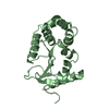
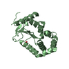
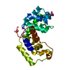
- PDB-1un2: Crystal structure of circularly permuted CPDSBA_Q100T99: Preserve... -
+ Open data

- Basic information
| Entry | Database: PDB / ID: 1un2 | |||||||||
|---|---|---|---|---|---|---|---|---|---|---|
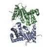
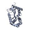
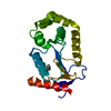
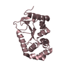
| Title | Crystal structure of circularly permuted CPDSBA_Q100T99: Preserved Global Fold and Local Structural Adjustments | |||||||||
 Components Components | THIOL-DISULFIDE INTERCHANGE PROTEIN | |||||||||
 Keywords Keywords |  OXIDOREDUCTASE / DISULFIDE OXIDOREDUCTASE / PROTEIN DISULFIDE ISOMERASE / PROTEIN FOLDING / THIOREDOXIN / REDOX PROTEIN / DISULFIDE BOND FORMATION / CIRCULAR PERMUTATION OXIDOREDUCTASE / DISULFIDE OXIDOREDUCTASE / PROTEIN DISULFIDE ISOMERASE / PROTEIN FOLDING / THIOREDOXIN / REDOX PROTEIN / DISULFIDE BOND FORMATION / CIRCULAR PERMUTATION | |||||||||
| Function / homology |  Function and homology information Function and homology informationcellular response to antibiotic / protein disulfide isomerase activity / protein-disulfide reductase activity / outer membrane-bounded periplasmic space / periplasmic space / oxidoreductase activitySimilarity search - Function | |||||||||
| Biological species |  ESCHERICHIA COLI (E. coli) ESCHERICHIA COLI (E. coli) | |||||||||
| Method | X-RAY DIFFRACTION / SYNCHROTRON / MOLECULAR REPLACEMENT / Resolution: 2.4 Å | |||||||||
 Authors Authors | Manjasetty, B.A. / Hennecke, J. / Glockshuber, R. / Heinemann, U. | |||||||||
 Citation Citation | Journal: Acta Crystallogr.,Sect.D / Year: 2004 Title: Structure of Circularly Permuted Dsba(Q100T99): Preserved Global Fold and Local Structural Adjustments Authors: Manjasetty, B.A. / Hennecke, J. / Glockshuber, R. / Heinemann, U. #1: Journal: J.Mol.Biol. / Year: 1999 Title: Random Circular Permutaion of Dsba Reveals Segments that are Essential for Protein Folding and Stability Authors: Hennecke, J. / Sebbal, P. / Glockshuber, R. | |||||||||
| History |
|
- Structure visualization
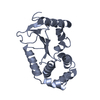
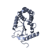
| Structure viewer | Molecule: MolmilJmol/JSmol |
|---|
- Downloads & links
-Download
| PDBx/mmCIF format | 1un2.cif.gz | 53.8 KB | Display | PDBx/mmCIF format |
|---|---|---|---|---|
| PDB format | pdb1un2.ent.gz | 37.7 KB | Display | PDB format |
| PDBx/mmJSON format | 1un2.json.gz | Tree view | PDBx/mmJSON format | |
| Others |  Other downloads Other downloads |
-Validation report
| Arichive directory | https://data.pdbj.org/pub/pdb/validation_reports/un/1un2ftp://data.pdbj.org/pub/pdb/validation_reports/un/1un2 | HTTPS FTP |
|---|
-Related structure data
| Related structure data | 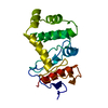 1a2jS S: Starting model for refinement |
|---|---|
| Similar structure data |


-Links
 PDBj
PDBj


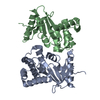
- Assembly
| Deposited unit | 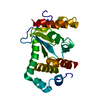
| ||||||||
|---|---|---|---|---|---|---|---|---|---|
| 1 |
| ||||||||
| Unit cell |
|
-Components
| #1: Protein | Mass: 21839.832 Da / Num. of mol.: 1 Fragment: THIOREDOXIN-LIKE DOMAIN, HELICAL DOMAIN RESIDUES, 119-208 Source method: isolated from a genetically manipulated source Details: DISULFIDE BOND FORMATION PROTEIN / Source: (gene. exp.) ESCHERICHIA COLI (E. coli) / Plasmid: PDSBA5 / Cellular location (production host): PERIPLASM / Production host: ESCHERICHIA COLI (E. coli) / Strain (production host): KPNI / References: UniProt: P24991, UniProt: P0AEG4*PLUS |
|---|---|
| #2: Water | ChemComp-HOH / Water Mass: 18.015 Da / Num. of mol.: 82 / Source method: isolated from a natural source / Formula: H2O Mass: 18.015 Da / Num. of mol.: 82 / Source method: isolated from a natural source / Formula: H2O |
| Compound details | CPDSBA_Q100T99 IS A CIRCULARLY PERMUTED VARIANT OF THE PARENT THIOL-DISULFIDE INTERCHANGE PROTEIN ...CPDSBA_Q100T99 IS A CIRCULARLY |
| Sequence details | THE PDB HAS CHANGED THE SEQUENCE NUMBERING IN THIS ENTRY FROM THAT USED IN THE PUBLISHED LITERATURE. ...THE PDB HAS CHANGED THE SEQUENCE NUMBERING IN THIS ENTRY FROM THAT USED IN THE PUBLISHED LITERATURE |
-Experimental details
-Experiment
| Experiment | Method: X-RAY DIFFRACTION / Number of used crystals: 1 |
|---|
- Sample preparation
| Crystal | Density Matthews: 2.2 Å3/Da / Density % sol: 45 % | ||||||||||||||||||||||||||||||
|---|---|---|---|---|---|---|---|---|---|---|---|---|---|---|---|---|---|---|---|---|---|---|---|---|---|---|---|---|---|---|---|
| Crystal grow | pH: 6.5 Details: 25% PEG 8000, 7-10% DMSO, 0.1M NA CACODYLATE PH 6.5 | ||||||||||||||||||||||||||||||
| Crystal grow | *PLUS Temperature: 291 K / pH: 6.5 / Method: vapor diffusion, hanging drop | ||||||||||||||||||||||||||||||
| Components of the solutions | *PLUS
|
-Data collection
| Diffraction | Mean temperature: 100 K |
|---|---|
| Diffraction source | Source: SYNCHROTRON / Site: EMBL/DESY, HAMBURG  / Beamline: X11 / Wavelength: 0.9116 / Beamline: X11 / Wavelength: 0.9116 |
| Detector | Date: Apr 8, 1999 |
| Radiation | Protocol: SINGLE WAVELENGTH / Monochromatic (M) / Laue (L): M / Scattering type: x-ray |
| Radiation wavelength | Wavelength: 0.9116 Å / Relative weight: 1 |
| Reflection | Resolution: 2.4→20 Å / Num. obs: 7952 / % possible obs: 99 % / Rmerge(I) obs: 0.056 / Net I/σ(I): 17.4 |
| Reflection shell | Resolution: 2.4→2.48 Å / Rmerge(I) obs: 0.099 / Mean I/σ(I) obs: 6.4 / % possible all: 98.1 |
| Reflection | *PLUS Highest resolution: 2.4 Å / Num. measured all: 37723 / Rmerge(I) obs: 0.056 |
| Reflection shell | *PLUS % possible obs: 98.1 % / Rmerge(I) obs: 0.099 / Mean I/σ(I) obs: 6.4 |
- Processing
| Software |
| ||||||||||||||||||||||||||||||||||||||||||||||||||||||||||||||||||||||||||||||||||||||||||||||||||||||||||||||||||||||||||||||||||||||||||||||||||||||||||||||||||||||||||||||||||||||
|---|---|---|---|---|---|---|---|---|---|---|---|---|---|---|---|---|---|---|---|---|---|---|---|---|---|---|---|---|---|---|---|---|---|---|---|---|---|---|---|---|---|---|---|---|---|---|---|---|---|---|---|---|---|---|---|---|---|---|---|---|---|---|---|---|---|---|---|---|---|---|---|---|---|---|---|---|---|---|---|---|---|---|---|---|---|---|---|---|---|---|---|---|---|---|---|---|---|---|---|---|---|---|---|---|---|---|---|---|---|---|---|---|---|---|---|---|---|---|---|---|---|---|---|---|---|---|---|---|---|---|---|---|---|---|---|---|---|---|---|---|---|---|---|---|---|---|---|---|---|---|---|---|---|---|---|---|---|---|---|---|---|---|---|---|---|---|---|---|---|---|---|---|---|---|---|---|---|---|---|---|---|---|---|
| Refinement | Method to determine structure: MOLECULAR REPLACEMENT Starting model: PDB ENTRY 1A2J Resolution: 2.4→20 Å / Cor.coef. Fo:Fc: 0.943 / Cor.coef. Fo:Fc free: 0.913 / SU B: 10.016 / SU ML: 0.224 / TLS residual ADP flag: LIKELY RESIDUAL / Cross valid method: THROUGHOUT / ESU R: 0.627 / ESU R Free: 0.297 / Stereochemistry target values: MAXIMUM LIKELIHOOD / Details: HYDROGENS HAVE BEEN ADDED IN THE RIDING POSITIONS
| ||||||||||||||||||||||||||||||||||||||||||||||||||||||||||||||||||||||||||||||||||||||||||||||||||||||||||||||||||||||||||||||||||||||||||||||||||||||||||||||||||||||||||||||||||||||
| Solvent computation | Ion probe radii: 0.8 Å / Shrinkage radii: 0.8 Å / VDW probe radii: 1.4 Å / Solvent model: BABINET MODEL WITH MASK | ||||||||||||||||||||||||||||||||||||||||||||||||||||||||||||||||||||||||||||||||||||||||||||||||||||||||||||||||||||||||||||||||||||||||||||||||||||||||||||||||||||||||||||||||||||||
| Displacement parameters | Biso mean: 38.61 Å2
| ||||||||||||||||||||||||||||||||||||||||||||||||||||||||||||||||||||||||||||||||||||||||||||||||||||||||||||||||||||||||||||||||||||||||||||||||||||||||||||||||||||||||||||||||||||||
| Refinement step | Cycle: LAST / Resolution: 2.4→20 Å
| ||||||||||||||||||||||||||||||||||||||||||||||||||||||||||||||||||||||||||||||||||||||||||||||||||||||||||||||||||||||||||||||||||||||||||||||||||||||||||||||||||||||||||||||||||||||
| Refine LS restraints |
|