 Movie
Movie Controller

[English] 日本語
 Yorodumi
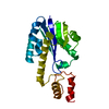
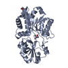
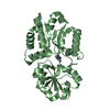
- PDB-1uax: Crystal structure of the ribonuclease H2 from Pyrococcus horikosh... -
+ Open data

- Basic information
| Entry | Database: PDB / ID: 1uax | ||||||
|---|---|---|---|---|---|---|---|
| Title | Crystal structure of the ribonuclease H2 from Pyrococcus horikoshii OT3 | ||||||
 Components Components | Ribonuclease HII | ||||||
 Keywords Keywords |  HYDROLASE / RNA*DNA hybrid ribonucleotidohydrolase HYDROLASE / RNA*DNA hybrid ribonucleotidohydrolase | ||||||
| Function / homology |  Function and homology information Function and homology informationRNA catabolic process / ribonuclease H / RNA-DNA hybrid ribonuclease activity / manganese ion binding / RNA binding / cytoplasmSimilarity search - Function | ||||||
| Biological species |   Pyrococcus horikoshii (archaea) Pyrococcus horikoshii (archaea) | ||||||
| Method | X-RAY DIFFRACTION / SYNCHROTRON / MOLECULAR REPLACEMENT / Resolution: 2 Å | ||||||
 Authors Authors | Hata, T. / Numata, T. / Kakuta, Y. / Kimura, M. | ||||||
 Citation Citation | Journal: To be published Title: Crystal structure of the ribonuclease H2 from Pyrococcus horikoshii OT3 Authors: Hata, T. / Numata, T. / Kakuta, Y. / Kimura, M. | ||||||
| History |
|
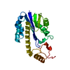
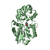
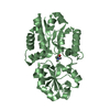
- Structure visualization
| Structure viewer | Molecule: MolmilJmol/JSmol |
|---|
- Downloads & links
-Download
| PDBx/mmCIF format | 1uax.cif.gz | 97.3 KB | Display | PDBx/mmCIF format |
|---|---|---|---|---|
| PDB format | pdb1uax.ent.gz | 75.4 KB | Display | PDB format |
| PDBx/mmJSON format | 1uax.json.gz | Tree view | PDBx/mmJSON format | |
| Others |  Other downloads Other downloads |
-Validation report
| Arichive directory | https://data.pdbj.org/pub/pdb/validation_reports/ua/1uaxftp://data.pdbj.org/pub/pdb/validation_reports/ua/1uax | HTTPS FTP |
|---|
-Related structure data
| Related structure data |  1io2S S: Starting model for refinement |
|---|---|
| Similar structure data |


-Links
 PDBj
PDBj
- Assembly
| Deposited unit | 
| ||||||||
|---|---|---|---|---|---|---|---|---|---|
| 1 | 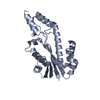
| ||||||||
| 2 | 
| ||||||||
| Unit cell |
|
-Components
| #1: Protein | Mass: 25326.160 Da / Num. of mol.: 2 Source method: isolated from a genetically manipulated source Source: (gene. exp.) Pyrococcus horikoshii (archaea) / Strain: OT3 / Plasmid: pET22b / Production host:  Escherichia coli (E. coli) / Strain (production host): BL21(DE3) codon plus RIL / References: UniProt: O59351, ribonuclease H Escherichia coli (E. coli) / Strain (production host): BL21(DE3) codon plus RIL / References: UniProt: O59351, ribonuclease H#2: Water | ChemComp-HOH / | Water Mass: 18.015 Da / Num. of mol.: 193 / Source method: isolated from a natural source / Formula: H2O Mass: 18.015 Da / Num. of mol.: 193 / Source method: isolated from a natural source / Formula: H2O |
|---|
-Experimental details
-Experiment
| Experiment | Method: X-RAY DIFFRACTION / Number of used crystals: 1 |
|---|
- Sample preparation
| Crystal | Density Matthews: 2.16 Å3/Da / Density % sol: 42.64 % |
|---|---|
| Crystal grow | Temperature: 293 K / Method: vapor diffusion, hanging drop / pH: 8.5 Details: 0.1M Tris-HCl, 2M Ammonium sulfate, pH 8.5, VAPOR DIFFUSION, HANGING DROP, temperature 293K |
-Data collection
| Diffraction | Mean temperature: 100 K |
|---|---|
| Diffraction source | Source: SYNCHROTRON / Site: SPring-8  / Beamline: BL44XU / Wavelength: 0.9 Å / Beamline: BL44XU / Wavelength: 0.9 Å |
| Detector | Type: OXFORD / Detector: CCD / Date: Feb 1, 2002 / Details: mirrors |
| Radiation | Monochromator: Si111 CHANNEL / Protocol: SINGLE WAVELENGTH / Monochromatic (M) / Laue (L): M / Scattering type: x-ray |
| Radiation wavelength | Wavelength: 0.9 Å / Relative weight: 1 |
| Reflection | Resolution: 2→41.65 Å / Num. all: 55176 / Num. obs: 55176 / % possible obs: 97.6 % / Observed criterion σ(F): 0 / Observed criterion σ(I): 0 / Redundancy: 3.39 % / Biso Wilson estimate: 8.7 Å2 / Rmerge(I) obs: 0.1 / Net I/σ(I): 12.2 |
| Reflection shell | Resolution: 2→2.07 Å / Redundancy: 3.3 % / Rmerge(I) obs: 0.126 / Mean I/σ(I) obs: 6.4 / Num. unique all: 2825 / % possible all: 95.6 |
- Processing
| Software |
| |||||||||||||||||||||||||
|---|---|---|---|---|---|---|---|---|---|---|---|---|---|---|---|---|---|---|---|---|---|---|---|---|---|---|
| Refinement | Method to determine structure: MOLECULAR REPLACEMENT Starting model: PDB ENTRY 1IO2 Resolution: 2→41.65 Å / Rfactor Rfree error: 0.006 / Data cutoff high absF: 486885.1 / Data cutoff low absF: 0 / Isotropic thermal model: RESTRAINED / Cross valid method: THROUGHOUT / σ(F): 0 / σ(I): 0 / Stereochemistry target values: Engh & Huber
| |||||||||||||||||||||||||
| Solvent computation | Solvent model: FLAT MODEL / Bsol: 44.8482 Å2 / ksol: 0.364108 e/Å3 | |||||||||||||||||||||||||
| Displacement parameters | Biso mean: 18.9 Å2
| |||||||||||||||||||||||||
| Refine analyze |
| |||||||||||||||||||||||||
| Refinement step | Cycle: LAST / Resolution: 2→41.65 Å
| |||||||||||||||||||||||||
| Refine LS restraints |
| |||||||||||||||||||||||||
| LS refinement shell | Resolution: 2→2.13 Å / Rfactor Rfree error: 0.015 / Total num. of bins used: 6
| |||||||||||||||||||||||||
| Xplor file |
|
