 Movie
Movie Controller

[English] 日本語
 Yorodumi
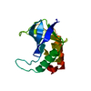
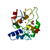
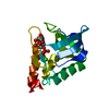
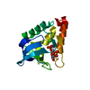
- PDB-1u9r: Crystal Structure of Staphylococcal Nuclease mutant V66E/P117G/H1... -
+ Open data

- Basic information
| Entry | Database: PDB / ID: 1u9r | ||||||
|---|---|---|---|---|---|---|---|
| Title | Crystal Structure of Staphylococcal Nuclease mutant V66E/P117G/H124L/S128A at Room Temperature | ||||||
 Components Components | Thermonuclease Micrococcal nuclease Micrococcal nuclease | ||||||
 Keywords Keywords | HYDROLASE / Staphylococcal nuclease / nuclease / hyperstable variant / internal waters | ||||||
| Function / homology |  Function and homology information Function and homology informationendonuclease activity, active with either ribo- or deoxyribonucleic acids and producing 3'-phosphomonoesters / micrococcal nuclease / nucleic acid binding / extracellular region / membrane / metal ion bindingSimilarity search - Function | ||||||
| Biological species |   Staphylococcus aureus (bacteria) Staphylococcus aureus (bacteria) | ||||||
| Method | X-RAY DIFFRACTION / MOLECULAR REPLACEMENT / Resolution: 2.1 Å | ||||||
 Authors Authors | Denisov, V.P. / Schlessman, J.L. / Garcia-Moreno, B.E. / Halle, B. | ||||||
 Citation Citation | Journal: Biophys.J. / Year: 2004 Title: Stabilization of internal charges in a protein: water penetration or conformational change? Authors: Denisov, V.P. / Schlessman, J.L. / Garcia-Moreno, B.E. / Halle, B. | ||||||
| History |
|
- Structure visualization
| Structure viewer | Molecule: MolmilJmol/JSmol |
|---|
- Downloads & links
-Download
| PDBx/mmCIF format | 1u9r.cif.gz | 37.8 KB | Display | PDBx/mmCIF format |
|---|---|---|---|---|
| PDB format | pdb1u9r.ent.gz | 26 KB | Display | PDB format |
| PDBx/mmJSON format | 1u9r.json.gz | Tree view | PDBx/mmJSON format | |
| Others |  Other downloads Other downloads |
-Validation report
| Arichive directory | https://data.pdbj.org/pub/pdb/validation_reports/u9/1u9rftp://data.pdbj.org/pub/pdb/validation_reports/u9/1u9r | HTTPS FTP |
|---|
-Related structure data
| Related structure data | |
|---|---|
| Similar structure data |


-Links
 PDBj
PDBj- Assembly
| Deposited unit | 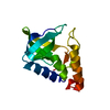
| ||||||||
|---|---|---|---|---|---|---|---|---|---|
| 1 |
| ||||||||
| Unit cell |
|
-Components
| #1: Protein | Micrococcal nuclease / TNase / Micrococcal nuclease / Staphylococcal nuclease Mass: 16792.260 Da / Num. of mol.: 1 / Mutation: V66E/P117G/H124L/S128A Source method: isolated from a genetically manipulated source Source: (gene. exp.) Staphylococcus aureus (bacteria) / Gene: nuc / Plasmid: lambda / Cell line (production host): AR120 / Production host: Escherichia coli (E. coli) / References: UniProt: P00644, micrococcal nuclease |
|---|---|
| #2: Water | ChemComp-HOH / Water Mass: 18.015 Da / Num. of mol.: 32 / Source method: isolated from a natural source / Formula: H2O Mass: 18.015 Da / Num. of mol.: 32 / Source method: isolated from a natural source / Formula: H2O |
-Experimental details
-Experiment
| Experiment | Method: X-RAY DIFFRACTION / Number of used crystals: 1 |
|---|
- Sample preparation
| Crystal | Density Matthews: 2.254 Å3/Da / Density % sol: 45.5 % |
|---|---|
| Crystal grow | Temperature: 277 K / Method: vapor diffusion, hanging drop / pH: 6.4 Details: MPD, potassium phosphate, pH 6.4, VAPOR DIFFUSION, HANGING DROP, temperature 277K |
-Data collection
| Diffraction | Mean temperature: 298 K |
|---|---|
| Diffraction source | Source: ROTATING ANODE / Type: RIGAKU RU300 / Wavelength: 1.5418 Å |
| Detector | Type: RIGAKU RAXIS IV / Detector: IMAGE PLATE / Date: Jul 1, 2003 / Details: mirrors |
| Radiation | Monochromator: yale mirrors / Protocol: SINGLE WAVELENGTH / Monochromatic (M) / Laue (L): M / Scattering type: x-ray |
| Radiation wavelength | Wavelength: 1.5418 Å / Relative weight: 1 |
| Reflection | Resolution: 2.1→50 Å / Num. all: 8569 / Num. obs: 8569 / % possible obs: 97.7 % / Observed criterion σ(F): 0 / Observed criterion σ(I): 0 / Redundancy: 4.5 % / Biso Wilson estimate: 20.3 Å2 / Rmerge(I) obs: 0.072 / Net I/σ(I): 28.8 |
| Reflection shell | Resolution: 2.1→2.18 Å / Redundancy: 4.5 % / Rmerge(I) obs: 0.215 / Mean I/σ(I) obs: 10 / Num. unique all: 826 / % possible all: 96 |
- Processing
| Software |
| ||||||||||||||||||||||||||||||||||||
|---|---|---|---|---|---|---|---|---|---|---|---|---|---|---|---|---|---|---|---|---|---|---|---|---|---|---|---|---|---|---|---|---|---|---|---|---|---|
| Refinement | Method to determine structure: MOLECULAR REPLACEMENT Starting model: Staphylococcal nuclease V66E/P117G/H124L/S128A low-temperature coordinates Resolution: 2.1→48.79 Å / Rfactor Rfree error: 0.007 / Data cutoff high absF: 519402.13 / Data cutoff low absF: 0 / Isotropic thermal model: RESTRAINED / Cross valid method: THROUGHOUT / σ(F): 0 / σ(I): 0 / Stereochemistry target values: Engh & Huber
| ||||||||||||||||||||||||||||||||||||
| Solvent computation | Solvent model: FLAT MODEL / Bsol: 52.7182 Å2 / ksol: 0.345061 e/Å3 | ||||||||||||||||||||||||||||||||||||
| Displacement parameters | Biso mean: 34.9 Å2
| ||||||||||||||||||||||||||||||||||||
| Refine analyze |
| ||||||||||||||||||||||||||||||||||||
| Refinement step | Cycle: LAST / Resolution: 2.1→48.79 Å
| ||||||||||||||||||||||||||||||||||||
| Refine LS restraints |
| ||||||||||||||||||||||||||||||||||||
| LS refinement shell | Resolution: 2.1→2.23 Å / Rfactor Rfree error: 0.023 / Total num. of bins used: 6
| ||||||||||||||||||||||||||||||||||||
| Xplor file |
|
