 Movie
Movie Controller


+ Open data

- Basic information
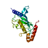
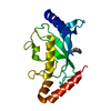
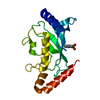
| Entry | Database: PDB / ID: 1u9b | ||||||
|---|---|---|---|---|---|---|---|
| Title | MURINE/HUMAN UBIQUITIN-CONJUGATING ENZYME UBC9 | ||||||
 Components Components | UBIQUITIN-CONJUGATING ENZYME E9 | ||||||
 Keywords Keywords |  LIGASE / UBIQUITIN-CONJUGATING ENZYME / UBIQUITIN-DIRECTED PROTEOLYSIS / CELL CYCLE CONTROL LIGASE / UBIQUITIN-CONJUGATING ENZYME / UBIQUITIN-DIRECTED PROTEOLYSIS / CELL CYCLE CONTROL | ||||||
| Function / homology |  Function and homology information Function and homology informationSUMO is transferred from E1 to E2 (UBE2I, UBC9) / Negative regulation of activity of TFAP2 (AP-2) family transcription factors / SUMOylation of nuclear envelope proteins / Vitamin D (calciferol) metabolism / Postmitotic nuclear pore complex (NPC) reformation / SUMOylation of ubiquitinylation proteins / SUMOylation of SUMOylation proteins / SUMOylation of transcription factors / SUMOylation of DNA replication proteins / SUMOylation of RNA binding proteins ...SUMO is transferred from E1 to E2 (UBE2I, UBC9) / Negative regulation of activity of TFAP2 (AP-2) family transcription factors / SUMOylation of nuclear envelope proteins / Vitamin D (calciferol) metabolism / Postmitotic nuclear pore complex (NPC) reformation / SUMOylation of ubiquitinylation proteins / SUMOylation of SUMOylation proteins / SUMOylation of transcription factors / SUMOylation of DNA replication proteins / SUMOylation of RNA binding proteins / SUMOylation of DNA methylation proteins / SUMOylation of DNA damage response and repair proteins / SUMOylation of transcription cofactors / SUMOylation of chromatin organization proteins / SUMOylation of intracellular receptors / SUMOylation of immune response proteins / HLH domain binding / SUMO conjugating enzyme activity / Formation of Incision Complex in GG-NER / RING-like zinc finger domain binding / Processing of DNA double-strand break ends / SUMO ligase complex / Recruitment and ATM-mediated phosphorylation of repair and signaling proteins at DNA double strand breaks / bHLH transcription factor binding / transferase complex / small protein activating enzyme binding / Transferases; Acyltransferases; Aminoacyltransferases / nuclear export / SUMO transferase activity / protein sumoylation / nuclear pore / ionotropic glutamate receptor binding / chromosome segregation / transcription coregulator binding / protein modification process / fibrillar center / PML body / ubiquitin-protein transferase activity / nuclear envelope / positive regulation of canonical NF-kappaB signal transduction / nuclear body / positive regulation of cell migration / cell division / negative regulation of DNA-templated transcription / dendrite / enzyme binding / negative regulation of transcription by RNA polymerase II / nucleoplasm / ATP binding / nucleus / cytosol / cytoplasmSimilarity search - Function | ||||||
| Biological species |  Mus musculus (house mouse) Mus musculus (house mouse) | ||||||
| Method | X-RAY DIFFRACTION / SYNCHROTRON / MOLECULAR REPLACEMENT / Resolution: 2 Å | ||||||
 Authors Authors | Tong, H. / Hateboer, G. / Perrakis, A. / Bernards, R. / Sixma, T.K. | ||||||
 Citation Citation | Journal: J.Biol.Chem. / Year: 1997 Title: Crystal structure of murine/human Ubc9 provides insight into the variability of the ubiquitin-conjugating system. Authors: Tong, H. / Hateboer, G. / Perrakis, A. / Bernards, R. / Sixma, T.K. | ||||||
| History |
|
- Structure visualization
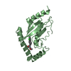
| Structure viewer | Molecule: MolmilJmol/JSmol |
|---|
- Downloads & links
-Download
| PDBx/mmCIF format | 1u9b.cif.gz | 45.9 KB | Display | PDBx/mmCIF format |
|---|---|---|---|---|
| PDB format | pdb1u9b.ent.gz | 32.2 KB | Display | PDB format |
| PDBx/mmJSON format | 1u9b.json.gz | Tree view | PDBx/mmJSON format | |
| Others |  Other downloads Other downloads |
-Validation report
| Arichive directory | https://data.pdbj.org/pub/pdb/validation_reports/u9/1u9bftp://data.pdbj.org/pub/pdb/validation_reports/u9/1u9b | HTTPS FTP |
|---|
-Related structure data
| Related structure data |  1u9aC  1aak S: Starting model for refinement C: citing same article ( |
|---|---|
| Similar structure data |


-Links
 PDBj
PDBj


- Assembly
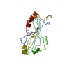
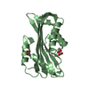
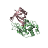
| Deposited unit | 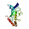
| ||||||||
|---|---|---|---|---|---|---|---|---|---|
| 1 |
| ||||||||
| Unit cell |
|
-Components
| #1: Protein | Mass: 18258.076 Da / Num. of mol.: 1 / Mutation: INS(ASN 0) Source method: isolated from a genetically manipulated source Details: MAMMALIAN / Source: (gene. exp.) Mus musculus (house mouse) / Production host:  Escherichia coli (E. coli) / Strain (production host): DH5ALPHA / References: UniProt: P63280, ubiquitin-protein ligase Escherichia coli (E. coli) / Strain (production host): DH5ALPHA / References: UniProt: P63280, ubiquitin-protein ligase |
|---|---|
| #2: Water | ChemComp-HOH / Water Mass: 18.015 Da / Num. of mol.: 96 / Source method: isolated from a natural source / Formula: H2O Mass: 18.015 Da / Num. of mol.: 96 / Source method: isolated from a natural source / Formula: H2O |
-Experimental details
-Experiment
| Experiment | Method: X-RAY DIFFRACTION / Number of used crystals: 1 |
|---|
- Sample preparation
| Crystal | Density Matthews: 2.64 Å3/Da / Density % sol: 52 % | ||||||||||||||||||||||||||||||||||||||||||||||||||||||||||||||||||||||||||||||
|---|---|---|---|---|---|---|---|---|---|---|---|---|---|---|---|---|---|---|---|---|---|---|---|---|---|---|---|---|---|---|---|---|---|---|---|---|---|---|---|---|---|---|---|---|---|---|---|---|---|---|---|---|---|---|---|---|---|---|---|---|---|---|---|---|---|---|---|---|---|---|---|---|---|---|---|---|---|---|---|
| Crystal grow | pH: 6.5 Details: 23% PEG MONOMETHYL ETHER 5000,9% ISOPROPANOL, 0.1 M AMMONIUM SULFATE, 0.1 M MES BUFFER, PH6.5 | ||||||||||||||||||||||||||||||||||||||||||||||||||||||||||||||||||||||||||||||
| Crystal grow | *PLUS Method: vapor diffusion, hanging drop | ||||||||||||||||||||||||||||||||||||||||||||||||||||||||||||||||||||||||||||||
| Components of the solutions | *PLUS
|
-Data collection
| Diffraction | Mean temperature: 281 K |
|---|---|
| Diffraction source | Source: SYNCHROTRON / Site: EMBL/DESY, HAMBURG  / Beamline: X11 / Wavelength: 0.885 / Beamline: X11 / Wavelength: 0.885 |
| Detector | Type: MARRESEARCH / Detector: IMAGE PLATE / Date: May 26, 1996 |
| Radiation | Monochromatic (M) / Laue (L): M / Scattering type: x-ray |
| Radiation wavelength | Wavelength: 0.885 Å / Relative weight: 1 |
| Reflection | Resolution: 2→20 Å / Num. obs: 12854 / % possible obs: 98 % / Observed criterion σ(I): 1 / Redundancy: 3.2 % / Biso Wilson estimate: 29.7 Å2 / Rmerge(I) obs: 0.058 / Rsym value: 0.058 / Net I/σ(I): 17 |
| Reflection shell | Resolution: 2.02→2.05 Å / Redundancy: 3.2 % / Rmerge(I) obs: 0.41 / Mean I/σ(I) obs: 3 / Rsym value: 0.41 / % possible all: 99 |
| Reflection shell | *PLUS % possible obs: 99 % |
- Processing
| Software |
| ||||||||||||||||||||||||||||||||||||||||||||||||||
|---|---|---|---|---|---|---|---|---|---|---|---|---|---|---|---|---|---|---|---|---|---|---|---|---|---|---|---|---|---|---|---|---|---|---|---|---|---|---|---|---|---|---|---|---|---|---|---|---|---|---|---|
| Refinement | Method to determine structure: MOLECULAR REPLACEMENT Starting model: PDB ENTRY 1AAK 1aak Resolution: 2→10 Å / Isotropic thermal model: TNT BCORREL V1.0 / σ(F): 0 / Stereochemistry target values: TNT PROTGEO
| ||||||||||||||||||||||||||||||||||||||||||||||||||
| Solvent computation | Solvent model: BABINET SCALING / Bsol: 228.3 Å2 / ksol: 0.8 e/Å3 | ||||||||||||||||||||||||||||||||||||||||||||||||||
| Refinement step | Cycle: LAST / Resolution: 2→10 Å
| ||||||||||||||||||||||||||||||||||||||||||||||||||
| Refine LS restraints |
| ||||||||||||||||||||||||||||||||||||||||||||||||||
| Software | *PLUS Name: TNT / Version: 5D / Classification: refinement | ||||||||||||||||||||||||||||||||||||||||||||||||||
| Refinement | *PLUS Rfactor obs: 0.185 / Rfactor Rfree: 0.252 | ||||||||||||||||||||||||||||||||||||||||||||||||||
| Solvent computation | *PLUS | ||||||||||||||||||||||||||||||||||||||||||||||||||
| Displacement parameters | *PLUS Biso mean: 26.1 Å2 | ||||||||||||||||||||||||||||||||||||||||||||||||||
| Refine LS restraints | *PLUS
|
