SEQUENCE According to the authors the final electron density maps clearly indicated that Gly167 and ...SEQUENCE According to the authors the final electron density maps clearly indicated that Gly167 and Asp357 were wrongly assigned in the SWS entry. The electron density was clear for Asp at position 167 and for GLY at position 357.
Mass: 18.015 Da / Num. of mol.: 2336 / Source method: isolated from a natural source / Formula: H2O
-
Experimental details
-
Experiment
Experiment
Method: X-RAY DIFFRACTION / Number of used crystals: 1
-
Sample preparation
Crystal
Density Matthews: 2.25 Å3/Da / Density % sol: 45 % Description: The number of unique observed reflections 563317 for the data collection and refinement refers to unmerged Friedel pairs. The value 296112 from the structure factor file refers to merged pairs.
Crystal grow
Temperature: 298 K / Method: vapor diffusion, sitting drop / pH: 8 Details: PEG 4000, lithium sulfate, pH 8, VAPOR DIFFUSION, SITTING DROP, temperature 298K
In the structure databanks used in Yorodumi, some data are registered as the other names, "COVID-19 virus" and "2019-nCoV". Here are the details of the virus and the list of structure data.
Jan 31, 2019. EMDB accession codes are about to change! (news from PDBe EMDB page)
EMDB accession codes are about to change! (news from PDBe EMDB page)
The allocation of 4 digits for EMDB accession codes will soon come to an end. Whilst these codes will remain in use, new EMDB accession codes will include an additional digit and will expand incrementally as the available range of codes is exhausted. The current 4-digit format prefixed with “EMD-” (i.e. EMD-XXXX) will advance to a 5-digit format (i.e. EMD-XXXXX), and so on. It is currently estimated that the 4-digit codes will be depleted around Spring 2019, at which point the 5-digit format will come into force.
The EM Navigator/Yorodumi systems omit the EMD- prefix.
Related info.:Q: What is EMD? / ID/Accession-code notation in Yorodumi/EM Navigator
Yorodumi is a browser for structure data from EMDB, PDB, SASBDB, etc.
This page is also the successor to EM Navigator detail page, and also detail information page/front-end page for Omokage search.
The word "yorodu" (or yorozu) is an old Japanese word meaning "ten thousand". "mi" (miru) is to see.
Related info.:EMDB / PDB / SASBDB / Comparison of 3 databanks / Yorodumi Search / Aug 31, 2016. New EM Navigator & Yorodumi / Yorodumi Papers / Jmol/JSmol / Function and homology information / Changes in new EM Navigator and Yorodumi
 Movie
Movie Controller

 Yorodumi
 Open data
 Basic information
 Components
 Keywords
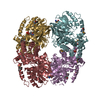
 LYASE /
  Function and homology information
 Clostridium aminobutyricum (bacteria)
 Authors
 Citation
 Structure visualization
 Downloads & links
 Other downloads


 PDBj
PDBj Assembly
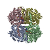

 Mass: 351.640 Da / Num. of mol.: 4 / Source method: obtained synthetically / Formula: Fe4S4
 Mass: 785.550 Da / Num. of mol.: 4 / Source method: obtained synthetically / Formula: C27H33N9O15P2 / Comment: FAD*YM
 Mass: 18.015 Da / Num. of mol.: 2336 / Source method: isolated from a natural source / Formula: H2O
 Sample preparation
 / Beamline: X06SA / Wavelength: 0.90004,1.73652,1.74314
 Processing