 Movie
Movie Controller

[English] 日本語
 Yorodumi
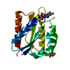
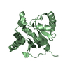
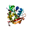
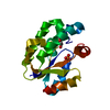
- PDB-1u8a: Crystal Structure of Mycobacterium Tuberculosis Shikimate Kinase ... -
+ Open data

- Basic information
| Entry | Database: PDB / ID: 1u8a | ||||||
|---|---|---|---|---|---|---|---|
| Title | Crystal Structure of Mycobacterium Tuberculosis Shikimate Kinase in Complex with Shikimate and ADP at 2.15 Angstrom Resolution | ||||||
 Components Components | Shikimate kinase | ||||||
 Keywords Keywords | TRANSFERASE / SHIKIMATE PATHWAY / SHIKIMATE KINASE / 2 PHORSPHORYL TRANSFER / DRUG DESIGN | ||||||
| Function / homology |  Function and homology informationshikimate kinase / shikimate metabolic process / Chorismate via Shikimate Pathway / shikimate kinase activity / chorismate biosynthetic process / aromatic amino acid family biosynthetic process / amino acid biosynthetic process / phosphorylation / magnesium ion binding / ATP binding / cytosol Function and homology informationshikimate kinase / shikimate metabolic process / Chorismate via Shikimate Pathway / shikimate kinase activity / chorismate biosynthetic process / aromatic amino acid family biosynthetic process / amino acid biosynthetic process / phosphorylation / magnesium ion binding / ATP binding / cytosolSimilarity search - Function | ||||||
| Biological species |   Mycobacterium tuberculosis (bacteria) Mycobacterium tuberculosis (bacteria) | ||||||
| Method | X-RAY DIFFRACTION / MOLECULAR REPLACEMENT / Resolution: 2.15 Å | ||||||
 Authors Authors | Dhaliwal, B. / Nichols, C.E. / Ren, J. / Lockyer, M. / Charles, I. / Hawkins, A.R. / Stammers, D.K. | ||||||
 Citation Citation | Journal: Febs Lett. / Year: 2004 Title: Crystallographic studies of shikimate binding and induced conformational changes in Mycobacterium tuberculosis shikimate kinase. Authors: Dhaliwal, B. / Nichols, C.E. / Ren, J. / Lockyer, M. / Charles, I. / Hawkins, A.R. / Stammers, D.K. | ||||||
| History |
|
- Structure visualization
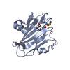
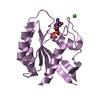
| Structure viewer | Molecule: MolmilJmol/JSmol |
|---|
- Downloads & links
-Download
| PDBx/mmCIF format | 1u8a.cif.gz | 48.3 KB | Display | PDBx/mmCIF format |
|---|---|---|---|---|
| PDB format | pdb1u8a.ent.gz | 33.2 KB | Display | PDB format |
| PDBx/mmJSON format | 1u8a.json.gz | Tree view | PDBx/mmJSON format | |
| Others |  Other downloads Other downloads |
-Validation report
| Arichive directory | https://data.pdbj.org/pub/pdb/validation_reports/u8/1u8aftp://data.pdbj.org/pub/pdb/validation_reports/u8/1u8a | HTTPS FTP |
|---|
-Related structure data
| Related structure data | 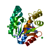 1l4yS S: Starting model for refinement |
|---|---|
| Similar structure data |


-Links
 PDBj
PDBj
- Assembly
| Deposited unit | 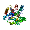
| ||||||||
|---|---|---|---|---|---|---|---|---|---|
| 1 |
| ||||||||
| Unit cell |
|
-Components
| #1: Protein | / SK Mass: 18612.352 Da / Num. of mol.: 1 Source method: isolated from a genetically manipulated source Source: (gene. exp.) Mycobacterium tuberculosis (bacteria) / Gene: aroK / Plasmid: pET17b / Species (production host): Escherichia coli / Production host: Escherichia coli BL21(DE3) (bacteria) / Strain (production host): BL21(DE3)References: UniProt: P0A4Z2, UniProt: P9WPY3*PLUS, shikimate kinase | ||||||
|---|---|---|---|---|---|---|---|
| #2: Chemical | Chloride  Mass: 35.453 Da / Num. of mol.: 3 / Source method: obtained synthetically / Formula: Cl Mass: 35.453 Da / Num. of mol.: 3 / Source method: obtained synthetically / Formula: Cl#3: Chemical | ChemComp-ADP / | Adenosine diphosphate  Mass: 427.201 Da / Num. of mol.: 1 / Source method: obtained synthetically / Formula: C10H15N5O10P2 / Comment: ADP, energy-carrying molecule*YM Mass: 427.201 Da / Num. of mol.: 1 / Source method: obtained synthetically / Formula: C10H15N5O10P2 / Comment: ADP, energy-carrying molecule*YM#4: Chemical | ChemComp-SKM / ( | Shikimic acid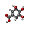  Mass: 174.151 Da / Num. of mol.: 1 / Source method: obtained synthetically / Formula: C7H10O5 Mass: 174.151 Da / Num. of mol.: 1 / Source method: obtained synthetically / Formula: C7H10O5#5: Water | ChemComp-HOH / | Water Mass: 18.015 Da / Num. of mol.: 117 / Source method: isolated from a natural source / Formula: H2O Mass: 18.015 Da / Num. of mol.: 117 / Source method: isolated from a natural source / Formula: H2O |
-Experimental details
-Experiment
| Experiment | Method: X-RAY DIFFRACTION / Number of used crystals: 1 |
|---|
- Sample preparation
| Crystal | Density Matthews: 2.79 Å3/Da / Density % sol: 55.53 % |
|---|---|
| Crystal grow | Temperature: 298 K / Method: vapor diffusion, sitting drop / pH: 7 Details: PEG 2000, Lithium chloride, Tris-HCl, Adenosine-5'-diphospate, Shikimic acid, pH 7, VAPOR DIFFUSION, SITTING DROP, temperature 298K |
-Data collection
| Diffraction | Mean temperature: 100 K |
|---|---|
| Diffraction source | Source: ROTATING ANODE / Type: RIGAKU RU200 / Wavelength: 1.5418 Å |
| Detector | Type: MARRESEARCH / Detector: IMAGE PLATE / Date: Feb 25, 2003 / Details: mirrors |
| Radiation | Monochromator: Osmic Confocal Blue Optics / Protocol: SINGLE WAVELENGTH / Monochromatic (M) / Laue (L): M / Scattering type: x-ray |
| Radiation wavelength | Wavelength: 1.5418 Å / Relative weight: 1 |
| Reflection | Resolution: 2.15→30 Å / Num. all: 12161 / Num. obs: 12135 / % possible obs: 99.9 % / Observed criterion σ(F): 0 / Redundancy: 5.93 % / Biso Wilson estimate: 40.52 Å2 / Rmerge(I) obs: 0.075 / Net I/σ(I): 22.4 |
| Reflection shell | Resolution: 2.15→2.19 Å / Redundancy: 5.86 % / Rmerge(I) obs: 0.644 / Mean I/σ(I) obs: 3.5 / Num. unique all: 606 / % possible all: 100 |
- Processing
| Software |
| |||||||||||||||||||||||||
|---|---|---|---|---|---|---|---|---|---|---|---|---|---|---|---|---|---|---|---|---|---|---|---|---|---|---|
| Refinement | Method to determine structure: MOLECULAR REPLACEMENT Starting model: PDB ENTRY 1L4Y Resolution: 2.15→30 Å / Cross valid method: THROUGHOUT / σ(F): 0 / Stereochemistry target values: Engh & Huber
| |||||||||||||||||||||||||
| Refine analyze |
| |||||||||||||||||||||||||
| Refinement step | Cycle: LAST / Resolution: 2.15→30 Å
| |||||||||||||||||||||||||
| Refine LS restraints |
|
