 Movie
Movie Controller

[English] 日本語
 Yorodumi
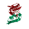
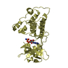
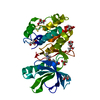
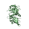
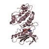
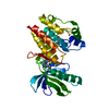
- PDB-1u73: Crystal structure of a Dimeric Acidic Platelet Aggregation Inhibi... -
+ Open data

- Basic information
| Entry | Database: PDB / ID: 1u73 | ||||||
|---|---|---|---|---|---|---|---|
| Title | Crystal structure of a Dimeric Acidic Platelet Aggregation Inhibitor and Hypotensive Phospholipase A2 from Bothrops jararacussu | ||||||
 Components Components | hypotensive phospholipase A2 | ||||||
 Keywords Keywords |  HYDROLASE / acidic phospholipase A2 / Bothrops jararacussu venom / platelet aggregation and hypotensive effects / oligomeric state / dimeric HYDROLASE / acidic phospholipase A2 / Bothrops jararacussu venom / platelet aggregation and hypotensive effects / oligomeric state / dimeric | ||||||
| Function / homology |  Function and homology informationphospholipase A2 activity / phospholipase A2 / arachidonic acid secretion / phospholipid metabolic process / lipid catabolic process / regulation of blood pressure / toxin activity / defense response to bacterium / calcium ion binding / extracellular region Function and homology informationphospholipase A2 activity / phospholipase A2 / arachidonic acid secretion / phospholipid metabolic process / lipid catabolic process / regulation of blood pressure / toxin activity / defense response to bacterium / calcium ion binding / extracellular regionSimilarity search - Function | ||||||
| Biological species |  Bothrops jararacussu (jararacussu) Bothrops jararacussu (jararacussu) | ||||||
| Method | X-RAY DIFFRACTION / SYNCHROTRON / MOLECULAR REPLACEMENT / Resolution: 1.9 Å | ||||||
 Authors Authors | Magro, A.J. / Murakami, M.T. / Soares, A.M. / Arni, R.K. / Fontes, M.R. | ||||||
 Citation Citation | Journal: Biochem.Biophys.Res.Commun. / Year: 2004 Title: Crystal structure of an acidic platelet aggregation inhibitor and hypotensive phospholipase A(2) in the monomeric and dimeric states: insights into its oligomeric state Authors: Magro, A.J. / Murakami, M.T. / Marcussi, S. / Soares, A.M. / Arni, R.K. / Fontes, M.R. #1: Journal: Biochem.Pharm. / Year: 2002Title: Structural and Functional Characterization of an Acidic Platelet Aggregation Inhibitor and Hypotensive Phospholipase A2 from Bothrops jararacussu Venom Authors: Andriao-Escarso, S.H. / Soares, A.M. / Fontes, M.R. / Fuly, A.L. / Correa, F.M. / Rosa, J.C. / Greene, L.J. / Giglio, J.R. | ||||||
| History |
|
- Structure visualization
| Structure viewer | Molecule: MolmilJmol/JSmol |
|---|
- Downloads & links
-Download
| PDBx/mmCIF format | 1u73.cif.gz | 66.4 KB | Display | PDBx/mmCIF format |
|---|---|---|---|---|
| PDB format | pdb1u73.ent.gz | 49.4 KB | Display | PDB format |
| PDBx/mmJSON format | 1u73.json.gz | Tree view | PDBx/mmJSON format | |
| Others |  Other downloads Other downloads |
-Validation report
| Arichive directory | https://data.pdbj.org/pub/pdb/validation_reports/u7/1u73ftp://data.pdbj.org/pub/pdb/validation_reports/u7/1u73 | HTTPS FTP |
|---|
-Related structure data


-Links
 PDBj
PDBj

- Assembly
| Deposited unit | 
| ||||||||
|---|---|---|---|---|---|---|---|---|---|
| 1 |
| ||||||||
| Unit cell |
|
-Components
| #1: Protein | Mass: 13700.451 Da / Num. of mol.: 2 / Source method: isolated from a natural source / Details: Venom glands / Source: (natural) Bothrops jararacussu (jararacussu) / References: UniProt: Q8AXY1, phospholipase A2#2: Water | ChemComp-HOH / | Water Mass: 18.015 Da / Num. of mol.: 381 / Source method: isolated from a natural source / Formula: H2O Mass: 18.015 Da / Num. of mol.: 381 / Source method: isolated from a natural source / Formula: H2O |
|---|
-Experimental details
-Experiment
| Experiment | Method: X-RAY DIFFRACTION / Number of used crystals: 1 |
|---|
- Sample preparation
| Crystal | Density Matthews: 1.733 Å3/Da / Density % sol: 29.04 % |
|---|---|
| Crystal grow | Temperature: 291 K / Method: vapor diffusion, hanging drop / pH: 3.5 Details: ammonium sulfate, PEG 6000, pH 3.5, VAPOR DIFFUSION, HANGING DROP, temperature 291K |
-Data collection
| Diffraction | Mean temperature: 100 K |
|---|---|
| Diffraction source | Source: SYNCHROTRON / Site: LNLS  / Beamline: D03B-MX1 / Wavelength: 1.38 Å / Beamline: D03B-MX1 / Wavelength: 1.38 Å |
| Detector | Type: MARRESEARCH / Detector: IMAGE PLATE / Date: Feb 15, 2002 / Details: Mirrors |
| Radiation | Monochromator: MIRRORS / Protocol: SINGLE WAVELENGTH / Monochromatic (M) / Laue (L): M / Scattering type: x-ray |
| Radiation wavelength | Wavelength: 1.38 Å / Relative weight: 1 |
| Reflection | Resolution: 1.9→29.7 Å / Num. all: 15160 / Num. obs: 14151 / % possible obs: 93.3 % / Observed criterion σ(F): 0 / Observed criterion σ(I): -3 / Redundancy: 3.5 % / Biso Wilson estimate: 24.8 Å2 / Rmerge(I) obs: 0.045 / Net I/σ(I): 20.1 |
| Reflection shell | Resolution: 1.9→2.02 Å / Redundancy: 3.4 % / Rmerge(I) obs: 0.293 / Mean I/σ(I) obs: 5 / Num. unique all: 2084 / % possible all: 87.7 |
- Processing
| Software |
| |||||||||||||||||||||||||
|---|---|---|---|---|---|---|---|---|---|---|---|---|---|---|---|---|---|---|---|---|---|---|---|---|---|---|
| Refinement | Method to determine structure: MOLECULAR REPLACEMENT Starting model: BthA-I (monomeric form) Resolution: 1.9→29.7 Å / Isotropic thermal model: Isotropic / Cross valid method: THROUGHOUT / σ(F): 0 / Stereochemistry target values: Engh & Huber
| |||||||||||||||||||||||||
| Displacement parameters | Biso mean: 27.8 Å2
| |||||||||||||||||||||||||
| Refine analyze | Luzzati coordinate error obs: 0.2 Å / Luzzati d res low obs: 4 Å / Luzzati sigma a obs: 0.22 Å | |||||||||||||||||||||||||
| Refinement step | Cycle: LAST / Resolution: 1.9→29.7 Å
| |||||||||||||||||||||||||
| Refine LS restraints |
|
