 Movie
Movie Controller

[English] 日本語
 Yorodumi
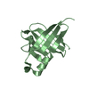
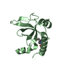
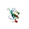
- PDB-1u5f: Crystal Structure of the PH Domain of SKAP-Hom with 8 Vector-deri... -
+ Open data

- Basic information
| Entry | Database: PDB / ID: 1u5f | ||||||
|---|---|---|---|---|---|---|---|
| Title | Crystal Structure of the PH Domain of SKAP-Hom with 8 Vector-derived N-terminal Residues | ||||||
 Components Components | Src-associated adaptor protein | ||||||
 Keywords Keywords |  SIGNALING PROTEIN / PH domain of SKAP-Hom / Artefactual dimerization induced by vector-derived sequence SIGNALING PROTEIN / PH domain of SKAP-Hom / Artefactual dimerization induced by vector-derived sequence | ||||||
| Function / homology |  Function and homology information Function and homology informationSignal regulatory protein family interactions / B cell activation / negative regulation of cell population proliferation / nucleoplasm / plasma membrane / cytosol / cytoplasmSimilarity search - Function | ||||||
| Biological species |  Mus musculus (house mouse) Mus musculus (house mouse) | ||||||
| Method | X-RAY DIFFRACTION / SYNCHROTRON / SIRAS / Resolution: 1.9 Å | ||||||
 Authors Authors | Tang, Y. / Swanson, K.D. / Neel, B.G. / Eck, M.J. | ||||||
 Citation Citation | Journal: To be Published Title: Structural Basis for the Dimerization and Phosphoinositide Specificity of the Src Kinase-associated Phosphoproteins SKAP55 and SKAP-Hom Authors: Tang, Y. / Swanson, K.D. / Neel, B.G. / Eck, M.J. | ||||||
| History |
| ||||||
| Remark 999 | SEQUENCE Thrombin cleavage results in a vector-derived linker peptide (RASVGSPG) before coding ...SEQUENCE Thrombin cleavage results in a vector-derived linker peptide (RASVGSPG) before coding sequence starting at residue 109. |
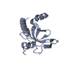
- Structure visualization
| Structure viewer | Molecule: MolmilJmol/JSmol |
|---|
- Downloads & links
-Download
| PDBx/mmCIF format | 1u5f.cif.gz | 38.1 KB | Display | PDBx/mmCIF format |
|---|---|---|---|---|
| PDB format | pdb1u5f.ent.gz | 26.4 KB | Display | PDB format |
| PDBx/mmJSON format | 1u5f.json.gz | Tree view | PDBx/mmJSON format | |
| Others |  Other downloads Other downloads |
-Validation report
| Arichive directory | https://data.pdbj.org/pub/pdb/validation_reports/u5/1u5fftp://data.pdbj.org/pub/pdb/validation_reports/u5/1u5f | HTTPS FTP |
|---|
-Related structure data


-Links
 PDBj
PDBj
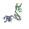
- Assembly
| Deposited unit | 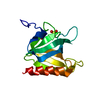
| ||||||||
|---|---|---|---|---|---|---|---|---|---|
| 1 |
| ||||||||
| 2 | 
| ||||||||
| Unit cell |
|
-Components
| #1: Protein | Mass: 17006.016 Da / Num. of mol.: 1 / Fragment: PH Domain, residues 109-248 / Mutation: A110P Source method: isolated from a genetically manipulated source Source: (gene. exp.) Mus musculus (house mouse) / Gene: SCAP2 / Plasmid: pGEX-2TK / Species (production host): Escherichia coli / Production host:  Escherichia coli BL21(DE3) (bacteria) / Strain (production host): BL21(DE3) / References: UniProt: Q8BK74, UniProt: Q3UND0*PLUS Escherichia coli BL21(DE3) (bacteria) / Strain (production host): BL21(DE3) / References: UniProt: Q8BK74, UniProt: Q3UND0*PLUS | ||
|---|---|---|---|
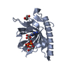
| #2: Chemical | Sulfate  Mass: 96.063 Da / Num. of mol.: 2 / Source method: obtained synthetically / Formula: SO4 Mass: 96.063 Da / Num. of mol.: 2 / Source method: obtained synthetically / Formula: SO4#3: Water | ChemComp-HOH / | Water Mass: 18.015 Da / Num. of mol.: 8 / Source method: isolated from a natural source / Formula: H2O Mass: 18.015 Da / Num. of mol.: 8 / Source method: isolated from a natural source / Formula: H2O |
-Experimental details
-Experiment
| Experiment | Method: X-RAY DIFFRACTION / Number of used crystals: 1 |
|---|
- Sample preparation
| Crystal | Density Matthews: 2.4 Å3/Da / Density % sol: 48.4 % |
|---|---|
| Crystal grow | Temperature: 298 K / Method: vapor diffusion, hanging drop / pH: 5.5 Details: Ammonium Sulfate, Sodium Citrate, pH 5.5, VAPOR DIFFUSION, HANGING DROP, temperature 298K |
-Data collection
| Diffraction | Mean temperature: 100 K |
|---|---|
| Diffraction source | Source: SYNCHROTRON / Site: CHESS  / Beamline: A1 / Wavelength: 0.916 Å / Beamline: A1 / Wavelength: 0.916 Å |
| Detector | Detector: CCD / Date: Jan 30, 2003 |
| Radiation | Monochromator: graphite / Protocol: SINGLE WAVELENGTH / Monochromatic (M) / Laue (L): M / Scattering type: x-ray |
| Radiation wavelength | Wavelength: 0.916 Å / Relative weight: 1 |
| Reflection | Resolution: 1.9→30 Å / Num. all: 12365 / Num. obs: 11889 / % possible obs: 95.2 % / Observed criterion σ(F): 0 / Observed criterion σ(I): 0 / Redundancy: 7.7 % / Biso Wilson estimate: 37 Å2 / Rmerge(I) obs: 0.072 / Net I/σ(I): 19.6 |
| Reflection shell | Resolution: 1.9→1.94 Å / Rmerge(I) obs: 0.429 / Mean I/σ(I) obs: 2 / Num. unique all: 797 / % possible all: 97.7 |
- Processing
| Software |
| ||||||||||||||||||||||||||||||||||||||||||||||||||||||||||||||||||||||||||||||||||||||||||||||||||||||||||||||||||||||||||||||||||||||||||||||||||||||||||||||||
|---|---|---|---|---|---|---|---|---|---|---|---|---|---|---|---|---|---|---|---|---|---|---|---|---|---|---|---|---|---|---|---|---|---|---|---|---|---|---|---|---|---|---|---|---|---|---|---|---|---|---|---|---|---|---|---|---|---|---|---|---|---|---|---|---|---|---|---|---|---|---|---|---|---|---|---|---|---|---|---|---|---|---|---|---|---|---|---|---|---|---|---|---|---|---|---|---|---|---|---|---|---|---|---|---|---|---|---|---|---|---|---|---|---|---|---|---|---|---|---|---|---|---|---|---|---|---|---|---|---|---|---|---|---|---|---|---|---|---|---|---|---|---|---|---|---|---|---|---|---|---|---|---|---|---|---|---|---|---|---|---|---|
| Refinement | Method to determine structure: SIRAS / Resolution: 1.9→30 Å / Cor.coef. Fo:Fc: 0.951 / Cor.coef. Fo:Fc free: 0.926 / SU B: 3.481 / SU ML: 0.102 / Cross valid method: THROUGHOUT / σ(F): 0 / σ(I): 0 / ESU R: 0.157 / ESU R Free: 0.139 / Stereochemistry target values: MAXIMUM LIKELIHOOD / Details: HYDROGENS HAVE BEEN ADDED IN THE RIDING POSITIONS
| ||||||||||||||||||||||||||||||||||||||||||||||||||||||||||||||||||||||||||||||||||||||||||||||||||||||||||||||||||||||||||||||||||||||||||||||||||||||||||||||||
| Solvent computation | Ion probe radii: 0.8 Å / Shrinkage radii: 0.8 Å / VDW probe radii: 1.4 Å / Solvent model: BABINET MODEL WITH MASK | ||||||||||||||||||||||||||||||||||||||||||||||||||||||||||||||||||||||||||||||||||||||||||||||||||||||||||||||||||||||||||||||||||||||||||||||||||||||||||||||||
| Displacement parameters | Biso mean: 48.919 Å2
| ||||||||||||||||||||||||||||||||||||||||||||||||||||||||||||||||||||||||||||||||||||||||||||||||||||||||||||||||||||||||||||||||||||||||||||||||||||||||||||||||
| Refinement step | Cycle: LAST / Resolution: 1.9→30 Å
| ||||||||||||||||||||||||||||||||||||||||||||||||||||||||||||||||||||||||||||||||||||||||||||||||||||||||||||||||||||||||||||||||||||||||||||||||||||||||||||||||
| Refine LS restraints |
| ||||||||||||||||||||||||||||||||||||||||||||||||||||||||||||||||||||||||||||||||||||||||||||||||||||||||||||||||||||||||||||||||||||||||||||||||||||||||||||||||
| LS refinement shell | Resolution: 1.901→1.95 Å / Total num. of bins used: 20
|
