 Movie
Movie Controller

[English] 日本語
 Yorodumi
- PDB-1u3h: Crystal structure of mouse TCR 172.10 complexed with MHC class II... -
+ Open data

- Basic information
| Entry | Database: PDB / ID: 1u3h | ||||||
|---|---|---|---|---|---|---|---|
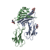
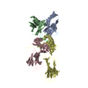
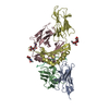
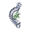
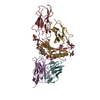
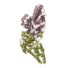
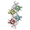
| Title | Crystal structure of mouse TCR 172.10 complexed with MHC class II I-Au molecule at 2.4 A | ||||||
 Components Components |
| ||||||
 Keywords Keywords |  IMMUNE SYSTEM / complex IMMUNE SYSTEM / complex | ||||||
| Function / homology |  Function and homology information Function and homology informationcompact myelin / structural constituent of myelin sheath / internode region of axon / negative regulation of axonogenesis / negative regulation of heterotypic cell-cell adhesion / antigen processing and presentation of peptide antigen / membrane organization / positive regulation of chemokine (C-X-C motif) ligand 2 production / positive regulation of T cell differentiation / T cell receptor complex ...compact myelin / structural constituent of myelin sheath / internode region of axon / negative regulation of axonogenesis / negative regulation of heterotypic cell-cell adhesion / antigen processing and presentation of peptide antigen / membrane organization / positive regulation of chemokine (C-X-C motif) ligand 2 production / positive regulation of T cell differentiation / T cell receptor complex / maintenance of blood-brain barrier / antigen processing and presentation / negative regulation of T cell proliferation / myelination / multivesicular body / response to progesterone / cell projection / cell periphery / sensory perception of sound / response to toxic substance / peptide antigen assembly with MHC class II protein complex / MHC class II protein complex / peptide antigen binding / positive regulation of interleukin-6 production / antigen processing and presentation of exogenous peptide antigen via MHC class II / positive regulation of immune response / MAPK cascade / positive regulation of T cell activation / myelin sheath / MHC class II protein complex binding / late endosome membrane / protease binding / adaptive immune response / lysosome / early endosome / cell surface receptor signaling pathway / calmodulin binding / lysosomal membrane / external side of plasma membrane / neuronal cell body / lipid binding / protein-containing complex binding / Golgi apparatus / cell surface / protein-containing complex / nucleus / plasma membrane / cytoplasmSimilarity search - Function | ||||||
| Biological species |  Mus musculus (house mouse) Mus musculus (house mouse)synthetic construct (others) | ||||||
| Method | X-RAY DIFFRACTION / SYNCHROTRON / MOLECULAR REPLACEMENT / Resolution: 2.42 Å | ||||||
 Authors Authors | Maynard, J. / Petersson, K. / Wilson, D.H. / Adams, E.J. / Blondelle, S.E. / Boulanger, M.J. / Wilson, D.B. / Garcia, K.C. | ||||||
 Citation Citation | Journal: Immunity / Year: 2005 Title: Structure of an autoimmune T cell receptor complexed with class II peptide-MHC: insights into MHC bias and antigen specificity Authors: Maynard, J. / Petersson, K. / Wilson, D.H. / Adams, E.J. / Blondelle, S.E. / Boulanger, M.J. / Wilson, D.B. / Garcia, K.C. | ||||||
| History |
|
- Structure visualization
| Structure viewer | Molecule: MolmilJmol/JSmol |
|---|
- Downloads & links
-Download
| PDBx/mmCIF format | 1u3h.cif.gz | 253.7 KB | Display | PDBx/mmCIF format |
|---|---|---|---|---|
| PDB format | pdb1u3h.ent.gz | 204.5 KB | Display | PDB format |
| PDBx/mmJSON format | 1u3h.json.gz | Tree view | PDBx/mmJSON format | |
| Others |  Other downloads Other downloads |
-Validation report
| Arichive directory | https://data.pdbj.org/pub/pdb/validation_reports/u3/1u3hftp://data.pdbj.org/pub/pdb/validation_reports/u3/1u3h | HTTPS FTP |
|---|
-Related structure data
| Related structure data | 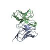 1d9kS S: Starting model for refinement |
|---|---|
| Similar structure data |


-Links
 PDBj
PDBj


- Assembly
| Deposited unit | 
| ||||||||
|---|---|---|---|---|---|---|---|---|---|
| 1 | 
| ||||||||
| 2 | 
| ||||||||
| Unit cell |
|
-Components
-Protein , 2 types, 4 molecules AEBF
| #1: Protein | Mass: 12290.634 Da / Num. of mol.: 2 / Fragment: V2.3-J39-C Source method: isolated from a genetically manipulated source Source: (gene. exp.) Mus musculus (house mouse) / Plasmid: pAK400 / Species (production host): Escherichia coli / Production host:  Escherichia coli BL21 (bacteria) / Strain (production host): BL21 / References: UniProt: Q5R1B3 Escherichia coli BL21 (bacteria) / Strain (production host): BL21 / References: UniProt: Q5R1B3#2: Protein | Mass: 12112.181 Da / Num. of mol.: 2 / Mutation: G17E,H47Y,I75T,L78S Source method: isolated from a genetically manipulated source Source: (gene. exp.) Mus musculus (house mouse) / Plasmid: pAK400 / Species (production host): Escherichia coli / Production host: Escherichia coli BL21 (bacteria) / Strain (production host): BL21 / References: UniProt: P04213 |
|---|
-H-2 class II histocompatibility antigen, A-U ... , 2 types, 4 molecules CGDH
| #3: Protein | Mass: 20670.047 Da / Num. of mol.: 2 / Fragment: extracellular alpha-1, extracellular alpha-2 Source method: isolated from a genetically manipulated source Source: (gene. exp.) Mus musculus (house mouse) / Gene: H2-Aa / Plasmid: pRMHa3 / Cell line (production host): S2 / Production host:  Drosophila melanogaster (fruit fly) / References: UniProt: P14438 Drosophila melanogaster (fruit fly) / References: UniProt: P14438#4: Protein | Mass: 22495.164 Da / Num. of mol.: 2 / Fragment: extracellular beta-1, extracellular beta-2 Source method: isolated from a genetically manipulated source Source: (gene. exp.) Mus musculus (house mouse) / Plasmid: pRMHa3 / Cell line (production host): S2 / Production host: Drosophila melanogaster (fruit fly) / References: UniProt: P06344 |
|---|
-Protein/peptide / Non-polymers , 2 types, 185 molecules PI
| #5: Protein/peptide | Mass: 1295.364 Da / Num. of mol.: 2 / Mutation: K4Y / Source method: obtained synthetically / Source: (synth.) synthetic construct (others) / References: UniProt: P04370*PLUS #6: Water | ChemComp-HOH / | WaterMass: 18.015 Da / Num. of mol.: 183 / Source method: isolated from a natural source / Formula: H2O |
|---|
-Experimental details
-Experiment
| Experiment | Method: X-RAY DIFFRACTION / Number of used crystals: 1 |
|---|
- Sample preparation
| Crystal | Density Matthews: 3.33 Å3/Da / Density % sol: 61.6 % |
|---|---|
| Crystal grow | Temperature: 291 K / Method: vapor diffusion, sitting drop / pH: 7.5 Details: 21% PEG 3350, 0.1M Hepes and 0.2M LiSO4, pH 7.5, VAPOR DIFFUSION, SITTING DROP, temperature 291K |
-Data collection
| Diffraction | Mean temperature: 100 K |
|---|---|
| Diffraction source | Source: SYNCHROTRON / Site: ALS  / Beamline: 8.2.1 / Wavelength: 1 Å / Beamline: 8.2.1 / Wavelength: 1 Å |
| Detector | Type: ADSC QUANTUM 4 / Detector: CCD / Date: Apr 3, 2004 |
| Radiation | Protocol: SINGLE WAVELENGTH / Monochromatic (M) / Laue (L): M / Scattering type: x-ray |
| Radiation wavelength | Wavelength: 1 Å / Relative weight: 1 |
| Reflection | Resolution: 2.4→50 Å / Num. all: 67849 / Num. obs: 67849 / % possible obs: 97.5 % / Observed criterion σ(F): 0 / Observed criterion σ(I): 0 / Redundancy: 9.2 % / Biso Wilson estimate: 37.7 Å2 / Rmerge(I) obs: 0.061 / Rsym value: 0.061 / Net I/σ(I): 20.2 |
| Reflection shell | Resolution: 2.4→2.49 Å / Redundancy: 10.5 % / Rmerge(I) obs: 0.386 / Mean I/σ(I) obs: 2.3 / Num. unique all: 6419 / Rsym value: 0.386 / % possible all: 93.5 |
- Processing
| Software |
| |||||||||||||||||||||||||
|---|---|---|---|---|---|---|---|---|---|---|---|---|---|---|---|---|---|---|---|---|---|---|---|---|---|---|
| Refinement | Method to determine structure: MOLECULAR REPLACEMENT Starting model: 1D9K Resolution: 2.42→41.51 Å / Rfactor Rfree error: 0.005 / Data cutoff high absF: 195797.18 / Data cutoff low absF: 0 / Isotropic thermal model: RESTRAINED / Cross valid method: THROUGHOUT / σ(F): -1 / σ(I): -1 / Stereochemistry target values: Engh & Huber
| |||||||||||||||||||||||||
| Solvent computation | Solvent model: FLAT MODEL / Bsol: 37.9538 Å2 / ksol: 0.330412 e/Å3 | |||||||||||||||||||||||||
| Displacement parameters | Biso mean: 61.7 Å2
| |||||||||||||||||||||||||
| Refine analyze |
| |||||||||||||||||||||||||
| Refinement step | Cycle: LAST / Resolution: 2.42→41.51 Å
| |||||||||||||||||||||||||
| Refine LS restraints |
| |||||||||||||||||||||||||
| LS refinement shell | Resolution: 2.4→2.55 Å / Rfactor Rfree error: 0.022 / Total num. of bins used: 6
| |||||||||||||||||||||||||
| Xplor file |
|
