 Movie
Movie Controller


+ Open data

- Basic information
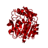
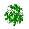
| Entry | Database: PDB / ID: 1u2e | ||||||
|---|---|---|---|---|---|---|---|
| Title | Crystal Structure of the C-C bond hydrolase MhpC | ||||||
 Components Components | 2-hydroxy-6-ketonona-2,4-dienedioic acid hydrolase | ||||||
 Keywords Keywords |  HYDROLASE / ALPHA/BETA HYDROLASE FOLD HYDROLASE / ALPHA/BETA HYDROLASE FOLD | ||||||
| Function / homology |  Function and homology information2-hydroxy-6-oxonona-2,4-dienedioate hydrolase / 2-hydroxy-6-oxonona-2,4,7-trienedioate hydrolase activity / 3-(3-hydroxy)phenylpropionate catabolic process / 2-hydroxy-6-oxonona-2,4-dienedioate hydrolase activity / 3-phenylpropionate catabolic process / : / hydrolase activity / protein homodimerization activity / cytoplasm Function and homology information2-hydroxy-6-oxonona-2,4-dienedioate hydrolase / 2-hydroxy-6-oxonona-2,4,7-trienedioate hydrolase activity / 3-(3-hydroxy)phenylpropionate catabolic process / 2-hydroxy-6-oxonona-2,4-dienedioate hydrolase activity / 3-phenylpropionate catabolic process / : / hydrolase activity / protein homodimerization activity / cytoplasmSimilarity search - Function | ||||||
| Biological species |  Escherichia coli (E. coli) Escherichia coli (E. coli) | ||||||
| Method | X-RAY DIFFRACTION / SYNCHROTRON / MOLECULAR REPLACEMENT / Resolution: 2.1 Å | ||||||
 Authors Authors | Montgomery, M.G. / Dunn, G. / Mohammed, F. / Robertson, T. / Garcia, J.-L. / Coker, A. / Bugg, T.D.H. / Wood, S.P. | ||||||
 Citation Citation | Journal: J.Mol.Biol. / Year: 2005 Title: The Structure of the C-C Bond Hydrolase MhpC Provides Insights into its Catalytic Mechanism Authors: Dunn, G. / Montgomery, M.G. / Mohammed, F. / Coker, A. / Cooper, J.B. / Robertson, T. / Garcia, J.-L. / Bugg, T.D.H. / Wood, S.P. | ||||||
| History |
|
- Structure visualization
| Structure viewer | Molecule: MolmilJmol/JSmol |
|---|
- Downloads & links
-Download
| PDBx/mmCIF format | 1u2e.cif.gz | 242.2 KB | Display | PDBx/mmCIF format |
|---|---|---|---|---|
| PDB format | pdb1u2e.ent.gz | 193.1 KB | Display | PDB format |
| PDBx/mmJSON format | 1u2e.json.gz | Tree view | PDBx/mmJSON format | |
| Others |  Other downloads Other downloads |
-Validation report
| Arichive directory | https://data.pdbj.org/pub/pdb/validation_reports/u2/1u2eftp://data.pdbj.org/pub/pdb/validation_reports/u2/1u2e | HTTPS FTP |
|---|
-Related structure data
| Similar structure data |
|---|


-Links
 PDBj
PDBj

- Assembly
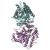
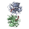
| Deposited unit | 
| ||||||||
|---|---|---|---|---|---|---|---|---|---|
| 1 | 
| ||||||||
| 2 | 
| ||||||||
| Unit cell |
|
-Components
| #1: Protein | Mass: 32105.631 Da / Num. of mol.: 4 Source method: isolated from a genetically manipulated source Source: (gene. exp.) Escherichia coli (E. coli) / Gene: mhpC / Plasmid: pIPC / Species (production host): Escherichia coli / Production host: Escherichia coli BL21(DE3) (bacteria) / Strain (production host): BL21(DE3) / References: UniProt: P77044, EC: 3.7.1.-#2: Chemical | ChemComp-CL / Chloride  Mass: 35.453 Da / Num. of mol.: 4 / Source method: obtained synthetically / Formula: Cl Mass: 35.453 Da / Num. of mol.: 4 / Source method: obtained synthetically / Formula: Cl#3: Water | ChemComp-HOH / | Water Mass: 18.015 Da / Num. of mol.: 544 / Source method: isolated from a natural source / Formula: H2O Mass: 18.015 Da / Num. of mol.: 544 / Source method: isolated from a natural source / Formula: H2O |
|---|
-Experimental details
-Experiment
| Experiment | Method: X-RAY DIFFRACTION / Number of used crystals: 1 |
|---|
- Sample preparation
| Crystal | Density Matthews: 2.59 Å3/Da / Density % sol: 53 % |
|---|---|
| Crystal grow | Temperature: 298 K / Method: vapor diffusion, hanging drop / pH: 8 Details: MPD, PEG 8000, Tris, Calcium chloride, pH 8.0, VAPOR DIFFUSION, HANGING DROP, temperature 298.0K |
-Data collection
| Diffraction | Mean temperature: 100 K |
|---|---|
| Diffraction source | Source: SYNCHROTRON / Site: SRS  / Beamline: PX14.2 / Wavelength: 0.87 Å / Beamline: PX14.2 / Wavelength: 0.87 Å |
| Detector | Type: ADSC QUANTUM 4 / Detector: CCD / Date: Oct 15, 2002 |
| Radiation | Monochromator: Si 111 CHANNEL / Protocol: SINGLE WAVELENGTH / Monochromatic (M) / Laue (L): M / Scattering type: x-ray |
| Radiation wavelength | Wavelength: 0.87 Å / Relative weight: 1 |
| Reflection | Resolution: 2.1→20 Å / Num. all: 73037 / Num. obs: 71688 / % possible obs: 93.7 % / Observed criterion σ(F): 0 / Observed criterion σ(I): 0 / Redundancy: 4.6 % / Biso Wilson estimate: 38.2 Å2 / Rmerge(I) obs: 0.075 / Net I/σ(I): 6.3 |
| Reflection shell | Resolution: 2.1→2.21 Å / Redundancy: 2 % / Rmerge(I) obs: 0.548 / Mean I/σ(I) obs: 1.3 / Num. unique all: 4674 / % possible all: 72.9 |
- Processing
| Software |
| |||||||||||||||||||||||||
|---|---|---|---|---|---|---|---|---|---|---|---|---|---|---|---|---|---|---|---|---|---|---|---|---|---|---|
| Refinement | Method to determine structure: MOLECULAR REPLACEMENT Starting model: MAD structure of MhpC Resolution: 2.1→20 Å / Isotropic thermal model: anisotropic / Cross valid method: Free-R / σ(F): 0 / Stereochemistry target values: Engh & Huber
| |||||||||||||||||||||||||
| Refinement step | Cycle: LAST / Resolution: 2.1→20 Å
| |||||||||||||||||||||||||
| Refine LS restraints |
|
