 Movie
Movie Controller

[English] 日本語
 Yorodumi
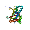
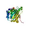
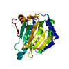
- PDB-1txo: Crystal structure of the Mycobacterium tuberculosis serine/threon... -
+ Open data

- Basic information
| Entry | Database: PDB / ID: 1txo | ||||||
|---|---|---|---|---|---|---|---|
| Title | Crystal structure of the Mycobacterium tuberculosis serine/threonine phosphatase PstP/Ppp at 1.95 A. | ||||||
 Components Components | Putative Bacterial Enzyme | ||||||
 Keywords Keywords |  HYDROLASE / Putative Bacterial Enzyme / Serine/threonine protein phosphatases / PstP/Ppp / Structural Genomics / PSI / Protein Structure Initiative / TB Structural Genomics Consortium / TBSGC HYDROLASE / Putative Bacterial Enzyme / Serine/threonine protein phosphatases / PstP/Ppp / Structural Genomics / PSI / Protein Structure Initiative / TB Structural Genomics Consortium / TBSGC | ||||||
| Function / homology |  Function and homology information Function and homology informationnegative regulation of catalytic activity / myosin phosphatase activity / protein serine/threonine phosphatase activity / protein-serine/threonine phosphatase / positive regulation of catalytic activity / negative regulation of DNA binding / phosphoprotein phosphatase activity / protein dephosphorylation / peptidoglycan-based cell wall / negative regulation of protein kinase activity ...negative regulation of catalytic activity / myosin phosphatase activity / protein serine/threonine phosphatase activity / protein-serine/threonine phosphatase / positive regulation of catalytic activity / negative regulation of DNA binding / phosphoprotein phosphatase activity / protein dephosphorylation / peptidoglycan-based cell wall / negative regulation of protein kinase activity / manganese ion binding / membrane => GO:0016020 / magnesium ion binding / membrane / metal ion binding / plasma membraneSimilarity search - Function | ||||||
| Biological species |   Mycobacterium tuberculosis (bacteria) Mycobacterium tuberculosis (bacteria) | ||||||
| Method | X-RAY DIFFRACTION / SYNCHROTRON / MAD / Resolution: 1.95 Å | ||||||
 Authors Authors | Pullen, K.E. / Ng, H.L. / Sung, P.Y. / Good, M.C. / Smith, S.M. / Alber, T. / TB Structural Genomics Consortium (TBSGC) | ||||||
 Citation Citation | Journal: Structure / Year: 2004 Title: An Alternate Conformation and a Third Metal in PstP/Ppp, the M. tuberculosis PP2C-Family Ser/Thr Protein Phosphatase. Authors: Pullen, K.E. / Ng, H.L. / Sung, P.Y. / Good, M.C. / Smith, S.M. / Alber, T. | ||||||
| History |
|
- Structure visualization
| Structure viewer | Molecule: MolmilJmol/JSmol |
|---|
- Downloads & links
-Download
| PDBx/mmCIF format | 1txo.cif.gz | 101.6 KB | Display | PDBx/mmCIF format |
|---|---|---|---|---|
| PDB format | pdb1txo.ent.gz | 82.3 KB | Display | PDB format |
| PDBx/mmJSON format | 1txo.json.gz | Tree view | PDBx/mmJSON format | |
| Others |  Other downloads Other downloads |
-Validation report
| Arichive directory | https://data.pdbj.org/pub/pdb/validation_reports/tx/1txoftp://data.pdbj.org/pub/pdb/validation_reports/tx/1txo | HTTPS FTP |
|---|
-Related structure data
| Similar structure data | |
|---|---|
| Other databases |


-Links
 PDBj
PDBj- Assembly
| Deposited unit | 
| ||||||||
|---|---|---|---|---|---|---|---|---|---|
| 1 | 
| ||||||||
| 2 | 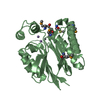
| ||||||||
| Unit cell |
|
-Components
| #1: Protein | Mass: 25483.650 Da / Num. of mol.: 2 Source method: isolated from a genetically manipulated source Source: (gene. exp.) Mycobacterium tuberculosis (bacteria) / Plasmid: pET24b / Production host: Escherichia coli (E. coli) / Strain (production host): BL21-CodonPlus (DE3)References: UniProt: Q8VKT2, UniProt: P9WHW5*PLUS, protein-serine/threonine phosphatase #2: Chemical | ChemComp-MN /   Mass: 54.938 Da / Num. of mol.: 6 / Source method: obtained synthetically / Formula: Mn Mass: 54.938 Da / Num. of mol.: 6 / Source method: obtained synthetically / Formula: Mn#3: Water | ChemComp-HOH / | Water Mass: 18.015 Da / Num. of mol.: 241 / Source method: isolated from a natural source / Formula: H2O Mass: 18.015 Da / Num. of mol.: 241 / Source method: isolated from a natural source / Formula: H2O |
|---|
-Experimental details
-Experiment
| Experiment | Method: X-RAY DIFFRACTION / Number of used crystals: 1 |
|---|
- Sample preparation
| Crystal | Density Matthews: 2.81 Å3/Da / Density % sol: 56.28 % |
|---|---|
| Crystal grow | Temperature: 291 K / Method: vapor diffusion, hanging drop / pH: 6 Details: PEG 10,000, sodium cacodylate, pH 6.0, VAPOR DIFFUSION, HANGING DROP, temperature 291K |
-Data collection
| Diffraction | Mean temperature: 100 K | |||||||||||||||
|---|---|---|---|---|---|---|---|---|---|---|---|---|---|---|---|---|
| Diffraction source | Source: SYNCHROTRON / Site: ALS  / Beamline: 8.3.1 / Wavelength: 1.12714, 0.97960, 0.95373, 1.8926 / Beamline: 8.3.1 / Wavelength: 1.12714, 0.97960, 0.95373, 1.8926 | |||||||||||||||
| Detector | Type: ADSC QUANTUM 4 / Detector: CCD / Date: Dec 6, 2003 | |||||||||||||||
| Radiation | Monochromator: DOUBLE CRYSTAL / Protocol: MAD / Monochromatic (M) / Laue (L): M / Scattering type: x-ray | |||||||||||||||
| Radiation wavelength |
| |||||||||||||||
| Reflection | Resolution: 1.95→72.55 Å / Num. all: 41602 / Num. obs: 38658 / % possible obs: 98 % / Observed criterion σ(F): 0 / Observed criterion σ(I): 0 / Redundancy: 3.5 % / Biso Wilson estimate: 35.6 Å2 / Rmerge(I) obs: 0.044 / Rsym value: 0.06 / Net I/σ(I): 18.3 | |||||||||||||||
| Reflection shell | Highest resolution: 1.95 Å / Redundancy: 3.5 % / Rmerge(I) obs: 0.352 / Mean I/σ(I) obs: 1.9 / Num. unique all: 41602 / Rsym value: 0.415 / % possible all: 98.1 |
- Processing
| Software |
| ||||||||||||||||||||||||||||||||||||||||||||||||||||||||||||||||||||||||||||||||||||||||||||||||||||||||||||||||||||||||||||||||||||||||||||||||||||||||||||||||
|---|---|---|---|---|---|---|---|---|---|---|---|---|---|---|---|---|---|---|---|---|---|---|---|---|---|---|---|---|---|---|---|---|---|---|---|---|---|---|---|---|---|---|---|---|---|---|---|---|---|---|---|---|---|---|---|---|---|---|---|---|---|---|---|---|---|---|---|---|---|---|---|---|---|---|---|---|---|---|---|---|---|---|---|---|---|---|---|---|---|---|---|---|---|---|---|---|---|---|---|---|---|---|---|---|---|---|---|---|---|---|---|---|---|---|---|---|---|---|---|---|---|---|---|---|---|---|---|---|---|---|---|---|---|---|---|---|---|---|---|---|---|---|---|---|---|---|---|---|---|---|---|---|---|---|---|---|---|---|---|---|---|
| Refinement | Method to determine structure: MAD / Resolution: 1.95→72.55 Å / Cor.coef. Fo:Fc: 0.951 / Cor.coef. Fo:Fc free: 0.933 / SU B: 3.631 / SU ML: 0.101 / Cross valid method: THROUGHOUT / σ(F): 0 / σ(I): 0 / ESU R: 0.157 / ESU R Free: 0.142 / Stereochemistry target values: MAXIMUM LIKELIHOOD / Details: HYDROGENS HAVE BEEN ADDED IN THE RIDING POSITIONS
| ||||||||||||||||||||||||||||||||||||||||||||||||||||||||||||||||||||||||||||||||||||||||||||||||||||||||||||||||||||||||||||||||||||||||||||||||||||||||||||||||
| Solvent computation | Ion probe radii: 0.8 Å / Shrinkage radii: 0.8 Å / VDW probe radii: 1.4 Å / Solvent model: BABINET MODEL WITH MASK | ||||||||||||||||||||||||||||||||||||||||||||||||||||||||||||||||||||||||||||||||||||||||||||||||||||||||||||||||||||||||||||||||||||||||||||||||||||||||||||||||
| Displacement parameters | Biso mean: 29.461 Å2
| ||||||||||||||||||||||||||||||||||||||||||||||||||||||||||||||||||||||||||||||||||||||||||||||||||||||||||||||||||||||||||||||||||||||||||||||||||||||||||||||||
| Refinement step | Cycle: LAST / Resolution: 1.95→72.55 Å
| ||||||||||||||||||||||||||||||||||||||||||||||||||||||||||||||||||||||||||||||||||||||||||||||||||||||||||||||||||||||||||||||||||||||||||||||||||||||||||||||||
| Refine LS restraints |
| ||||||||||||||||||||||||||||||||||||||||||||||||||||||||||||||||||||||||||||||||||||||||||||||||||||||||||||||||||||||||||||||||||||||||||||||||||||||||||||||||
| LS refinement shell | Resolution: 1.95→2.001 Å / Total num. of bins used: 20 /
|
