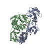
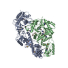
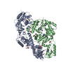
 Movie
Movie Controller


+ Open data

- Basic information
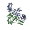
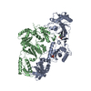
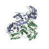
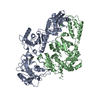
| Entry | Database: PDB / ID: 1tvr | ||||||
|---|---|---|---|---|---|---|---|
| Title | HIV-1 RT/9-CL TIBO | ||||||
 Components Components | (REVERSE TRANSCRIPTASE ) x 2 ) x 2 | ||||||
 Keywords Keywords | ASPARTYL PROTEASE / AIDS / POLYPROTEIN / HYDROLASE / ENDONUCLEASE / RNA-DIRECTED DNA POLYMERASE / 3HIV-1 RT/9-CL TIBO | ||||||
| Function / homology |  Function and homology information Function and homology information: / : / HIV-1 retropepsin / retroviral ribonuclease H / exoribonuclease H / exoribonuclease H activity / host multivesicular body / DNA integration / RNA-directed DNA polymerase / viral genome integration into host DNA ...: / : / HIV-1 retropepsin / retroviral ribonuclease H / exoribonuclease H / exoribonuclease H activity / host multivesicular body / DNA integration / RNA-directed DNA polymerase / viral genome integration into host DNA / viral penetration into host nucleus / establishment of integrated proviral latency / RNA-directed DNA polymerase activity / RNA-DNA hybrid ribonuclease activity / Transferases; Transferring phosphorus-containing groups; Nucleotidyltransferases / symbiont-mediated suppression of host gene expression / viral nucleocapsid / DNA recombination / Hydrolases; Acting on ester bonds / DNA-directed DNA polymerase / aspartic-type endopeptidase activity / DNA-directed DNA polymerase activity / symbiont entry into host cell / lipid binding / host cell nucleus / host cell plasma membrane / virion membrane / structural molecule activity / proteolysis / DNA binding / RNA binding / zinc ion binding / membraneSimilarity search - Function | ||||||
| Biological species |   Human immunodeficiency virus type 1 Human immunodeficiency virus type 1 | ||||||
| Method | X-RAY DIFFRACTION / SYNCHROTRON / Resolution: 3 Å | ||||||
 Authors Authors | Das, K. / Ding, J. / Hsiou, Y. / Arnold, E. | ||||||
 Citation Citation | Journal: J.Mol.Biol. / Year: 1996 Title: Crystal structures of 8-Cl and 9-Cl TIBO complexed with wild-type HIV-1 RT and 8-Cl TIBO complexed with the Tyr181Cys HIV-1 RT drug-resistant mutant. Authors: Das, K. / Ding, J. / Hsiou, Y. / Clark Jr., A.D. / Moereels, H. / Koymans, L. / Andries, K. / Pauwels, R. / Janssen, P.A. / Boyer, P.L. / Clark, P. / Smith Jr., R.H. / Kroeger Smith, M.B. / ...Authors: Das, K. / Ding, J. / Hsiou, Y. / Clark Jr., A.D. / Moereels, H. / Koymans, L. / Andries, K. / Pauwels, R. / Janssen, P.A. / Boyer, P.L. / Clark, P. / Smith Jr., R.H. / Kroeger Smith, M.B. / Michejda, C.J. / Hughes, S.H. / Arnold, E. #1: Journal: Drug Des.Discovery / Year: 1996Title: Targeting HIV Reverse Transcriptase for Anti-Aids Drug Design: Structural and Biological Considerations for Chemotherapeutic Strategies Authors: Arnold, E. / Das, K. / Ding, J. / Yadav, P.N. / Hsiou, Y. / Boyer, P.L. / Hughes, S.H. #2: Journal: Structure / Year: 1996Title: Structure of Unliganded HIV-1 Reverse Transcriptase at 2.7 A Resolution: Implications of Conformational Changes for Polymerization and Inhibition Mechanisms Authors: Hsiou, Y. / Ding, J. / Das, K. / Clark Junior, A.D. / Hughes, S.H. / Arnold, E. #3: Journal: Structure / Year: 1995Title: Structure of HIV-1 Reverse Transcriptase in a Complex with the Non-Nucleoside Inhibitor Alpha-Apa R 95845 at 2.8 A Resolution Authors: Ding, J. / Das, K. / Tantillo, C. / Zhang, W. / Clark Junior, A.D. / Jessen, S. / Lu, X. / Hsiou, Y. / Jacobo-Molina, A. / Andries, K. / al., et #4: Journal: Nat.Struct.Biol. / Year: 1995Title: Structure of HIV-1 RT/TIBO R 86183 Complex Reveals Similarity in the Binding of Diverse Nonnucleoside Inhibitors Authors: Ding, J. / Das, K. / Moereels, H. / Koymans, L. / Andries, K. / Janssen, P.A. / Hughes, S.H. / Arnold, E. #5: Journal: J.Mol.Biol. / Year: 1994Title: Locations of Anti-Aids Drug Binding Sites and Resistance Mutations in the Three-Dimensional Structure of HIV-1 Reverse Transcriptase. Implications for Mechanisms of Drug Inhibition and Resistance Authors: Tantillo, C. / Ding, J. / Jacobo-Molina, A. / Nanni, R.G. / Boyer, P.L. / Hughes, S.H. / Pauwels, R. / Andries, K. / Janssen, P.A. / Arnold, E. #6: Journal: J.Mol.Recog. / Year: 1994Title: Buried Surface Analysis of HIV-1 Reverse Transcriptase P66/P51 Heterodimer and its Interaction with DsDNA Template/Primer Authors: Ding, J. / Jacobo-Molina, A. / Tantillo, C. / Lu, X. / Nanni, R.G. / Arnold, E. #7: Journal: Proc.Natl.Acad.Sci.USA / Year: 1993Title: Crystal Structure of Human Immunodeficiency Virus Type 1 Reverse Transcriptase Complexed with Double-Stranded DNA at 3.0 A Resolution Shows Bent DNA Authors: Jacobo-Molina, A. / Ding, J. / Nanni, R.G. / Clark Junior, A.D. / Lu, X. / Tantillo, C. / Williams, R.L. / Kamer, G. / Ferris, A.L. / Clark, P. / al., et | ||||||
| History |
|
- Structure visualization
| Structure viewer | Molecule: MolmilJmol/JSmol |
|---|
- Downloads & links
-Download
| PDBx/mmCIF format | 1tvr.cif.gz | 204.6 KB | Display | PDBx/mmCIF format |
|---|---|---|---|---|
| PDB format | pdb1tvr.ent.gz | 162.4 KB | Display | PDB format |
| PDBx/mmJSON format | 1tvr.json.gz | Tree view | PDBx/mmJSON format | |
| Others |  Other downloads Other downloads |
-Validation report
| Arichive directory | https://data.pdbj.org/pub/pdb/validation_reports/tv/1tvrftp://data.pdbj.org/pub/pdb/validation_reports/tv/1tvr | HTTPS FTP |
|---|
-Related structure data


-Links
 PDBj
PDBj


- Assembly
| Deposited unit | 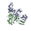
| ||||||||
|---|---|---|---|---|---|---|---|---|---|
| 1 |
| ||||||||
| Unit cell |
|
-Components
| #1: Protein | Mass: 64274.652 Da / Num. of mol.: 1 / Mutation: C280S Source method: isolated from a genetically manipulated source Source: (gene. exp.) Human immunodeficiency virus type 1 (CLONE 12)Genus: Lentivirus / Species: Human immunodeficiency virus 1Subtypes of HIV / Strain: BH10Description: HUMAN IMMUNODEFICIENCY VIRUS TYPE 1 (HIV-1) SUBCLONED FROM BH10 ISOLATE. EXPRESSED AND PROCESSED BY BACTERIAL ESCHERICHIA COLI. REF. A.HIZI, ET AL., (1988) PROC. NATL. ACAD. SCI. USA 85, ...Description: HUMAN IMMUNODEFICIENCY VIRUS TYPE 1 (HIV-1) SUBCLONED FROM BH10 ISOLATE. EXPRESSED AND PROCESSED BY BACTERIAL ESCHERICHIA COLI. REF. A.HIZI, ET AL., (1988) PROC. NATL. ACAD. SCI. USA 85, 1218-1222. REF. P.K.CLARK, ET AL., (1990) AIDS RES. HUM. RETROVIRUSES 6, 753-764. Cell line: 293 / Production host:  Escherichia coli (E. coli) / Strain (production host): 293 / References: UniProt: P03366, RNA-directed DNA polymerase Escherichia coli (E. coli) / Strain (production host): 293 / References: UniProt: P03366, RNA-directed DNA polymerase |
|---|---|
| #2: Protein | Mass: 49911.359 Da / Num. of mol.: 1 / Mutation: C280S Source method: isolated from a genetically manipulated source Source: (gene. exp.) Human immunodeficiency virus type 1 (CLONE 12)Genus: Lentivirus / Species: Human immunodeficiency virus 1Subtypes of HIV / Strain: BH10Description: HUMAN IMMUNODEFICIENCY VIRUS TYPE 1 (HIV-1) SUBCLONED FROM BH10 ISOLATE. EXPRESSED AND PROCESSED BY BACTERIAL ESCHERICHIA COLI. REF. A.HIZI, ET AL., (1988) PROC. NATL. ACAD. SCI. USA 85, ...Description: HUMAN IMMUNODEFICIENCY VIRUS TYPE 1 (HIV-1) SUBCLONED FROM BH10 ISOLATE. EXPRESSED AND PROCESSED BY BACTERIAL ESCHERICHIA COLI. REF. A.HIZI, ET AL., (1988) PROC. NATL. ACAD. SCI. USA 85, 1218-1222. REF. P.K.CLARK, ET AL., (1990) AIDS RES. HUM. RETROVIRUSES 6, 753-764. Cell line: 293 / Production host: Escherichia coli (E. coli) / Strain (production host): 293 / References: UniProt: P03366, RNA-directed DNA polymerase |
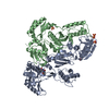
| #3: Chemical | ChemComp-TB9 / 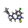  Mass: 321.868 Da / Num. of mol.: 1 / Source method: obtained synthetically / Formula: C16H20ClN3S Mass: 321.868 Da / Num. of mol.: 1 / Source method: obtained synthetically / Formula: C16H20ClN3S |
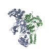
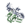
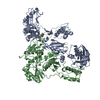
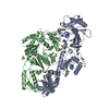
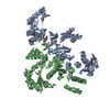
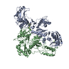
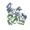
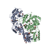
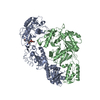
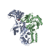
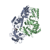
| Compound details | HIV-1 RT IS COMPOSED OF TWO SUBUNITS OF 66 KDA AND 51 KDA, DESIGNATED AS P66 AND P51, RESPECTIVELY. ...HIV-1 RT IS COMPOSED OF TWO SUBUNITS OF 66 KDA AND 51 KDA, DESIGNATED |
-Experimental details
-Experiment
| Experiment | Method: X-RAY DIFFRACTION |
|---|
- Sample preparation
| Crystal | Density Matthews: 3.41 Å3/Da / Density % sol: 65 % | ||||||||||||||||||||||||||||||||||||||||
|---|---|---|---|---|---|---|---|---|---|---|---|---|---|---|---|---|---|---|---|---|---|---|---|---|---|---|---|---|---|---|---|---|---|---|---|---|---|---|---|---|---|
| Crystal grow | *PLUS Temperature: 4 ℃ / pH: 6.8 / Method: vapor diffusion, hanging drop / Details: Clark, A.D., (1995) Methods Enzymol., 262, 171. | ||||||||||||||||||||||||||||||||||||||||
| Components of the solutions | *PLUS
|
-Data collection
| Diffraction source | Source: SYNCHROTRON / Site: CHESS  / Beamline: F1 / Wavelength: 0.918 / Beamline: F1 / Wavelength: 0.918 |
|---|---|
| Detector | Detector: CCD / Date: Nov 1, 1994 |
| Radiation | Monochromatic (M) / Laue (L): M / Scattering type: x-ray |
| Radiation wavelength | Wavelength: 0.918 Å / Relative weight: 1 |
| Reflection | Num. obs: 26209 / % possible obs: 84 % / Observed criterion σ(I): 1 / Redundancy: 2.16 % / Rmerge(I) obs: 0.064 |
| Reflection | *PLUS Highest resolution: 3 Å / Lowest resolution: 40 Å / Num. measured all: 56695 |
- Processing
| Software |
| ||||||||||||||||||||||||||||||||||||||||||||||||||||||||||||
|---|---|---|---|---|---|---|---|---|---|---|---|---|---|---|---|---|---|---|---|---|---|---|---|---|---|---|---|---|---|---|---|---|---|---|---|---|---|---|---|---|---|---|---|---|---|---|---|---|---|---|---|---|---|---|---|---|---|---|---|---|---|
| Refinement | Resolution: 3→10 Å / σ(F): 2 /
| ||||||||||||||||||||||||||||||||||||||||||||||||||||||||||||
| Refine analyze | Luzzati coordinate error obs: 0.6 Å | ||||||||||||||||||||||||||||||||||||||||||||||||||||||||||||
| Refinement step | Cycle: LAST / Resolution: 3→10 Å
| ||||||||||||||||||||||||||||||||||||||||||||||||||||||||||||
| Refine LS restraints |
| ||||||||||||||||||||||||||||||||||||||||||||||||||||||||||||
| Software | *PLUS Name: X-PLOR / Classification: refinement | ||||||||||||||||||||||||||||||||||||||||||||||||||||||||||||
| Refinement | *PLUS | ||||||||||||||||||||||||||||||||||||||||||||||||||||||||||||
| Solvent computation | *PLUS | ||||||||||||||||||||||||||||||||||||||||||||||||||||||||||||
| Displacement parameters | *PLUS |
