 Movie
Movie Controller


+ Open data

- Basic information
| Entry | Database: PDB / ID: 1tv2 | ||||||
|---|---|---|---|---|---|---|---|
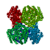
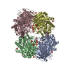
| Title | Crystal structure of the hydroxylamine MtmB complex | ||||||
 Components Components | Monomethylamine methyltransferase mtmB1 | ||||||
 Keywords Keywords |  TRANSFERASE / Tim barrel TRANSFERASE / Tim barrel | ||||||
| Function / homology |  Function and homology informationmethylamine-corrinoid protein Co-methyltransferase / monomethylamine methyltransferase activity / methanogenesis / methylation Function and homology informationmethylamine-corrinoid protein Co-methyltransferase / monomethylamine methyltransferase activity / methanogenesis / methylationSimilarity search - Function | ||||||
| Biological species |  Methanosarcina barkeri (archaea) Methanosarcina barkeri (archaea) | ||||||
| Method | X-RAY DIFFRACTION / SYNCHROTRON / FOURIER SYNTHESIS / Resolution: 2 Å | ||||||
 Authors Authors | Hao, B. / Zhao, G. / Kang, P.T. / Soares, J.A. / Ferguson, T.K. / Gallucci, J. / Krzycki, J.A. / Chan, M.K. | ||||||
 Citation Citation | Journal: Chem.Biol. / Year: 2004 Title: Reactivity and chemical synthesis of L-pyrrolysine- the 22(nd) genetically encoded amino acid Authors: Hao, B. / Zhao, G. / Kang, P.T. / Soares, J.A. / Ferguson, T.K. / Gallucci, J. / Krzycki, J.A. / Chan, M.K. | ||||||
| History |
|
- Structure visualization
| Structure viewer | Molecule: MolmilJmol/JSmol |
|---|
- Downloads & links
-Download
| PDBx/mmCIF format | 1tv2.cif.gz | 111.2 KB | Display | PDBx/mmCIF format |
|---|---|---|---|---|
| PDB format | pdb1tv2.ent.gz | 83.6 KB | Display | PDB format |
| PDBx/mmJSON format | 1tv2.json.gz | Tree view | PDBx/mmJSON format | |
| Others |  Other downloads Other downloads |
-Validation report
| Arichive directory | https://data.pdbj.org/pub/pdb/validation_reports/tv/1tv2ftp://data.pdbj.org/pub/pdb/validation_reports/tv/1tv2 | HTTPS FTP |
|---|
-Related structure data
| Related structure data |  1tv3C  1tv4C  1nthS S: Starting model for refinement C: citing same article ( |
|---|---|
| Similar structure data |


-Links
 PDBj
PDBj- Assembly
| Deposited unit | 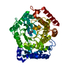
| ||||||||||||
|---|---|---|---|---|---|---|---|---|---|---|---|---|---|
| 1 | x 6
| ||||||||||||
| Unit cell |
| ||||||||||||
| Components on special symmetry positions |
|
-Components
| #1: Protein | Mass: 50183.117 Da / Num. of mol.: 1 / Source method: isolated from a natural source / Source: (natural) Methanosarcina barkeri (archaea) / Strain: MSReferences: UniProt: O30642, Transferases; Transferring one-carbon groups; Methyltransferases |
|---|---|
| #2: Chemical | ChemComp-BG5 /   Mass: 160.171 Da / Num. of mol.: 1 / Source method: obtained synthetically / Formula: C6H12N2O3 Mass: 160.171 Da / Num. of mol.: 1 / Source method: obtained synthetically / Formula: C6H12N2O3 |
| #3: Water | ChemComp-HOH / Water Mass: 18.015 Da / Num. of mol.: 461 / Source method: isolated from a natural source / Formula: H2O Mass: 18.015 Da / Num. of mol.: 461 / Source method: isolated from a natural source / Formula: H2O |
-Experimental details
-Experiment
| Experiment | Method: X-RAY DIFFRACTION / Number of used crystals: 1 |
|---|
- Sample preparation
| Crystal | Density Matthews: 4.29 Å3/Da / Density % sol: 71.1 % |
|---|---|
| Crystal grow | Temperature: 277 K / Method: vapor diffusion, hanging drop / pH: 7.5 Details: NACL, HEPES, pH 7.50, VAPOR DIFFUSION, HANGING DROP, temperature 277K |
-Data collection
| Diffraction | Mean temperature: 100 K |
|---|---|
| Diffraction source | Source: SYNCHROTRON / Site: SSRL  / Beamline: BL9-1 / Wavelength: 0.992 Å / Beamline: BL9-1 / Wavelength: 0.992 Å |
| Detector | Type: MARRESEARCH / Detector: CCD / Date: May 11, 2002 / Details: mirrors |
| Radiation | Monochromator: GRAPHITE / Protocol: SINGLE WAVELENGTH / Monochromatic (M) / Laue (L): M / Scattering type: x-ray |
| Radiation wavelength | Wavelength: 0.992 Å / Relative weight: 1 |
| Reflection | Resolution: 2→20 Å / Num. all: 67189 / Num. obs: 67189 / % possible obs: 98.9 % / Observed criterion σ(F): 0 / Observed criterion σ(I): 0 / Redundancy: 6.9 % / Biso Wilson estimate: 16.4 Å2 / Rmerge(I) obs: 0.177 / Rsym value: 0.177 / Net I/σ(I): 11.1 |
| Reflection shell | Resolution: 2→2.03 Å / Redundancy: 6.1 % / Rmerge(I) obs: 0.593 / Mean I/σ(I) obs: 2.2 / Num. unique all: 3307 / Rsym value: 0.593 / % possible all: 99.6 |
- Processing
| Software |
| |||||||||||||||||||||||||
|---|---|---|---|---|---|---|---|---|---|---|---|---|---|---|---|---|---|---|---|---|---|---|---|---|---|---|
| Refinement | Method to determine structure: FOURIER SYNTHESIS Starting model: 1NTH Resolution: 2→19.97 Å / Rfactor Rfree error: 0.002 / Data cutoff high absF: 311870.26 / Data cutoff low absF: 0 / Isotropic thermal model: RESTRAINED / Cross valid method: THROUGHOUT / σ(F): 0 / σ(I): 0 / Stereochemistry target values: Engh & Huber
| |||||||||||||||||||||||||
| Solvent computation | Solvent model: FLAT MODEL / Bsol: 65.3925 Å2 / ksol: 0.395048 e/Å3 | |||||||||||||||||||||||||
| Displacement parameters | Biso mean: 23.8 Å2
| |||||||||||||||||||||||||
| Refine analyze |
| |||||||||||||||||||||||||
| Refinement step | Cycle: LAST / Resolution: 2→19.97 Å
| |||||||||||||||||||||||||
| Refine LS restraints |
| |||||||||||||||||||||||||
| LS refinement shell | Resolution: 2→2.13 Å / Rfactor Rfree error: 0.008 / Total num. of bins used: 6
| |||||||||||||||||||||||||
| Xplor file |
|
