 Movie
Movie Controller

[English] 日本語
 Yorodumi
- PDB-1tv0: Solution structure of cryptdin-4, the most potent alpha-defensin ... -
+ Open data

- Basic information
| Entry | Database: PDB / ID: 1tv0 | ||||||
|---|---|---|---|---|---|---|---|
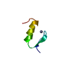
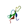
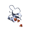
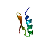
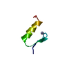
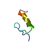
| Title | Solution structure of cryptdin-4, the most potent alpha-defensin from mouse Paneth cells | ||||||
 Components Components | Cryptdin-4 | ||||||
 Keywords Keywords |  ANTIMICROBIAL PROTEIN / beta sheet / beta hairpin ANTIMICROBIAL PROTEIN / beta sheet / beta hairpin | ||||||
| Function / homology |  Function and homology information Function and homology informationpore-forming activity / disruption of plasma membrane integrity in another organism / azurophil granule / transport vesicle / innate immune response in mucosa / phospholipid binding / antimicrobial humoral immune response mediated by antimicrobial peptide / antibacterial humoral response / killing of cells of another organism / cellular response to lipopolysaccharide ...pore-forming activity / disruption of plasma membrane integrity in another organism / azurophil granule / transport vesicle / innate immune response in mucosa / phospholipid binding / antimicrobial humoral immune response mediated by antimicrobial peptide / antibacterial humoral response / killing of cells of another organism / cellular response to lipopolysaccharide / defense response to Gram-negative bacterium / defense response to Gram-positive bacterium / defense response to bacterium / protein homodimerization activity / extracellular spaceSimilarity search - Function | ||||||
| Biological species |  Mus musculus (house mouse) Mus musculus (house mouse) | ||||||
| Method | SOLUTION NMR | ||||||
 Authors Authors | Jing, W. / Hunter, H.N. / Tanabe, H. / Ouellette, A.J. / Vogel, H.J. | ||||||
 Citation Citation | Journal: Biochemistry / Year: 2004 Title: Solution Structure of Cryptdin-4, a Mouse Paneth Cell alpha-Defensin. Authors: Jing, W. / Hunter, H.N. / Tanabe, H. / Ouellette, A.J. / Vogel, H.J. | ||||||
| History |
|
- Structure visualization
| Structure viewer | Molecule: MolmilJmol/JSmol |
|---|
- Downloads & links
-Download
| PDBx/mmCIF format | 1tv0.cif.gz | 203.7 KB | Display | PDBx/mmCIF format |
|---|---|---|---|---|
| PDB format | pdb1tv0.ent.gz | 177.3 KB | Display | PDB format |
| PDBx/mmJSON format | 1tv0.json.gz | Tree view | PDBx/mmJSON format | |
| Others |  Other downloads Other downloads |
-Validation report
| Arichive directory | https://data.pdbj.org/pub/pdb/validation_reports/tv/1tv0ftp://data.pdbj.org/pub/pdb/validation_reports/tv/1tv0 | HTTPS FTP |
|---|
-Related structure data
| Similar structure data |
|---|


-Links
 PDBj
PDBj
- Assembly
| Deposited unit | 
| |||||||||
|---|---|---|---|---|---|---|---|---|---|---|
| 1 |
| |||||||||
| NMR ensembles |
|
-Components
| #1: Protein/peptide | Mass: 3771.609 Da / Num. of mol.: 1 Source method: isolated from a genetically manipulated source Source: (gene. exp.) Mus musculus (house mouse) / Gene: DEFCR4 / Cell line (production host): BL21(DE3)-codon-Plus-RIL / Production host:  Escherichia coli (E. coli) / References: UniProt: P28311 Escherichia coli (E. coli) / References: UniProt: P28311 |
|---|
-Experimental details
-Experiment
| Experiment | Method: SOLUTION NMR | ||||||||||||||||
|---|---|---|---|---|---|---|---|---|---|---|---|---|---|---|---|---|---|
| NMR experiment |
| ||||||||||||||||
| NMR details | Text: BRUCKER MODEL EQUIPPED WITH TRIPPLE RESONANCE PROBE |
- Sample preparation
| Details | Contents: 3.2 mg protein H2O/D2O / Solvent system: H2O/D2O |
|---|---|
| Sample conditions | pH: 4.2 / Temperature: 303 K |
-NMR measurement
| Radiation | Protocol: SINGLE WAVELENGTH / Monochromatic (M) / Laue (L): M |
|---|---|
| Radiation wavelength | Relative weight: 1 |
| NMR spectrometer | Type: Bruker Bruker Advance 700 / Manufacturer: Bruker / Model: Bruker Advance 700 / Field strength: 700 MHz |
- Processing
| NMR software |
| ||||||||||||
|---|---|---|---|---|---|---|---|---|---|---|---|---|---|
| NMR representative | Selection criteria: fewest violations | ||||||||||||
| NMR ensemble | Conformers calculated total number: 100 / Conformers submitted total number: 20 |
