 Movie
Movie Controller

[English] 日本語
 Yorodumi
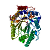
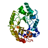
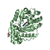
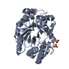
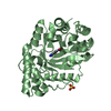
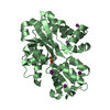
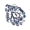
- PDB-1tux: HIGH RESOLUTION CRYSTAL STRUCTURE OF A THERMOSTABLE XYLANASE FROM... -
+ Open data

- Basic information
| Entry | Database: PDB / ID: 1tux | ||||||
|---|---|---|---|---|---|---|---|
| Title | HIGH RESOLUTION CRYSTAL STRUCTURE OF A THERMOSTABLE XYLANASE FROM THERMOASCUS AURANTIACUS | ||||||
 Components Components | XYLANASE | ||||||
 Keywords Keywords | HYDROLASE / XYLAN DEGRADATION / GLYCOSIDASE / ENZYME / 1 / 4-BETA-XYLAN XYLANOHYDROLASE | ||||||
| Function / homology |  Function and homology informationendo-1,4-beta-xylanase activity / endo-1,4-beta-xylanase / xylan catabolic process Function and homology informationendo-1,4-beta-xylanase activity / endo-1,4-beta-xylanase / xylan catabolic processSimilarity search - Function | ||||||
| Biological species |  Thermoascus aurantiacus (fungus) Thermoascus aurantiacus (fungus) | ||||||
| Method | X-RAY DIFFRACTION / MOLECULAR REPLACEMENT / Resolution: 1.8 Å | ||||||
 Authors Authors | Natesh, R. / Bhanumoorthy, P. / Vithayathil, P.J. / Sekar, K. / Ramakumar, S. / Viswamitra, M.A. | ||||||
 Citation Citation | Journal: J.Mol.Biol. / Year: 1999 Title: Crystal structure at 1.8 A resolution and proposed amino acid sequence of a thermostable xylanase from Thermoascus aurantiacus. Authors: Natesh, R. / Bhanumoorthy, P. / Vithayathil, P.J. / Sekar, K. / Ramakumar, S. / Viswamitra, M.A. #1: Journal: J.Mol.Biol. / Year: 1993Title: Crystallization and Preliminary X-Ray Diffraction Analysis of Crystals of Thermoascus Aurantiacus Xylanase Authors: Viswamitra, M.A. / Bhanumoorthy, P. / Ramakumar, S. / Manjula, M.V. / Vithayathil, P.J. / Murthy, S.K. / Naren, A.P. | ||||||
| History |
|
- Structure visualization
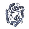
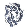
| Structure viewer | Molecule: MolmilJmol/JSmol |
|---|
- Downloads & links
-Download
| PDBx/mmCIF format | 1tux.cif.gz | 74.4 KB | Display | PDBx/mmCIF format |
|---|---|---|---|---|
| PDB format | pdb1tux.ent.gz | 54.5 KB | Display | PDB format |
| PDBx/mmJSON format | 1tux.json.gz | Tree view | PDBx/mmJSON format | |
| Others |  Other downloads Other downloads |
-Validation report
| Arichive directory | https://data.pdbj.org/pub/pdb/validation_reports/tu/1tuxftp://data.pdbj.org/pub/pdb/validation_reports/tu/1tux | HTTPS FTP |
|---|
-Related structure data
| Similar structure data |
|---|

-Links
 PDBj
PDBj
- Assembly
| Deposited unit | 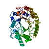
| ||||||||
|---|---|---|---|---|---|---|---|---|---|
| 1 |
| ||||||||
| Unit cell |
|
-Components
| #1: Protein | Mass: 32556.385 Da / Num. of mol.: 1 / Source method: isolated from a natural source Details: FUNGAL SOURCE FROM THERMOASCUS AURANTIACUS XYLANASE Source: (natural) Thermoascus aurantiacus (fungus) / Strain: LOCAL INDIAN SOIL / References: UniProt: P23360, endo-1,4-beta-xylanase |
|---|---|
| #2: Water | ChemComp-HOH / Water Mass: 18.015 Da / Num. of mol.: 266 / Source method: isolated from a natural source / Formula: H2O Mass: 18.015 Da / Num. of mol.: 266 / Source method: isolated from a natural source / Formula: H2O |
| Sequence details | THE SEQUENCE DEPOSITED IN THE SEQRES RECORDS IS IDENTIFIED |
-Experimental details
-Experiment
| Experiment | Method: X-RAY DIFFRACTION / Number of used crystals: 1 |
|---|
- Sample preparation
| Crystal | Density Matthews: 2.06 Å3/Da / Density % sol: 42 % Description: THE FULL ATOMIC COORDINATES OF THE WILD TYPE STREPTOMYCES LIVIDANS XYLANASE KINDLY PROVIDED TO US BY PROF. Z. DEREWENDA WERE USED AS THE STARTING MODEL FOR MOLECULAR REPLACEMENT. ONLY CA ...Description: THE FULL ATOMIC COORDINATES OF THE WILD TYPE STREPTOMYCES LIVIDANS XYLANASE KINDLY PROVIDED TO US BY PROF. Z. DEREWENDA WERE USED AS THE STARTING MODEL FOR MOLECULAR REPLACEMENT. ONLY CA COORDINATES OF STREPTOMYCES LIVIDANS XYLANASE ARE DEPOSITED IN PDB WITH ID CODE 1XAS TILL DATE. STRUCTURE REFINEMENT USING HIGH RESOLUTION DATA SETS AT 1.11 AND 0.89 A COLLECTED AT DIFFERENT TEMPERATURES IS CURRENTLY | ||||||||||||||||||||
|---|---|---|---|---|---|---|---|---|---|---|---|---|---|---|---|---|---|---|---|---|---|
| Crystal grow | pH: 7.2 / Details: pH 7.2 | ||||||||||||||||||||
| Crystal grow | *PLUS Method: vapor diffusion, hanging drop | ||||||||||||||||||||
| Components of the solutions | *PLUS
|
-Data collection
| Diffraction | Mean temperature: 293 K |
|---|---|
| Diffraction source | Source: ROTATING ANODE / Type: RIGAKU RUH2R / Wavelength: 1.5418 |
| Detector | Type: MARRESEARCH / Detector: IMAGE PLATE / Date: May 5, 1997 |
| Radiation | Monochromator: GRAPHITE(002) / Monochromatic (M) / Laue (L): M / Scattering type: x-ray |
| Radiation wavelength | Wavelength: 1.5418 Å / Relative weight: 1 |
| Reflection | Resolution: 1.8→99 Å / Num. obs: 22983 / % possible obs: 93 % / Redundancy: 3.7 % / Rmerge(I) obs: 0.05 |
| Reflection shell | Resolution: 1.8→1.86 Å / Rmerge(I) obs: 0.11 / % possible all: 86.1 |
| Reflection | *PLUS Num. measured all: 85248 |
| Reflection shell | *PLUS % possible obs: 86.1 % |
- Processing
| Software |
| ||||||||||||||||||||||||||||||||||||||||||||||||||||||||||||
|---|---|---|---|---|---|---|---|---|---|---|---|---|---|---|---|---|---|---|---|---|---|---|---|---|---|---|---|---|---|---|---|---|---|---|---|---|---|---|---|---|---|---|---|---|---|---|---|---|---|---|---|---|---|---|---|---|---|---|---|---|---|
| Refinement | Method to determine structure: MOLECULAR REPLACEMENT Starting model: STREPTOMYCES LIVIDANS XYLANASE Resolution: 1.8→10 Å / Rfactor Rfree error: 0.21 / Data cutoff high absF: 1000000 / Data cutoff low absF: 0.001 / Cross valid method: THROUGHOUT / σ(F): 2
| ||||||||||||||||||||||||||||||||||||||||||||||||||||||||||||
| Refine analyze |
| ||||||||||||||||||||||||||||||||||||||||||||||||||||||||||||
| Refinement step | Cycle: LAST / Resolution: 1.8→10 Å
| ||||||||||||||||||||||||||||||||||||||||||||||||||||||||||||
| Refine LS restraints |
| ||||||||||||||||||||||||||||||||||||||||||||||||||||||||||||
| LS refinement shell | Resolution: 1.8→1.86 Å / Total num. of bins used: 10
| ||||||||||||||||||||||||||||||||||||||||||||||||||||||||||||
| Xplor file |
| ||||||||||||||||||||||||||||||||||||||||||||||||||||||||||||
| Software | *PLUS Name: X-PLOR / Version: 3.851 / Classification: refinement | ||||||||||||||||||||||||||||||||||||||||||||||||||||||||||||
| Refine LS restraints | *PLUS
|
