 Movie
Movie Controller


+ Open data

- Basic information
| Entry | Database: PDB / ID: 1tpl | ||||||
|---|---|---|---|---|---|---|---|
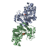
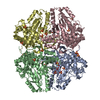
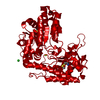
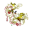
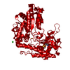
| Title | THE THREE-DIMENSIONAL STRUCTURE OF TYROSINE PHENOL-LYASE | ||||||
 Components Components | TYROSINE PHENOL-LYASE | ||||||
 Keywords Keywords | LYASE(CARBON-CARBON) | ||||||
| Function / homology |  Function and homology informationtyrosine phenol-lyase / tyrosine phenol-lyase activity / tyrosine metabolic process Function and homology informationtyrosine phenol-lyase / tyrosine phenol-lyase activity / tyrosine metabolic processSimilarity search - Function | ||||||
| Biological species |  Citrobacter intermedius (bacteria) Citrobacter intermedius (bacteria) | ||||||
| Method | X-RAY DIFFRACTION / Resolution: 2.3 Å | ||||||
 Authors Authors | Antson, A. / Demidkina, T. / Dauter, Z. / Harutyunyan, E. / Wilson, K. | ||||||
 Citation Citation | Journal: Biochemistry / Year: 1993 Title: Three-dimensional structure of tyrosine phenol-lyase. Authors: Antson, A.A. / Demidkina, T.V. / Gollnick, P. / Dauter, Z. / von Tersch, R.L. / Long, J. / Berezhnoy, S.N. / Phillips, R.S. / Harutyunyan, E.H. / Wilson, K.S. #1: Journal: FEBS Lett. / Year: 1992Title: The Polypeptide Chain Fold in Tyrosine Phenol-Lyase, a Pyridoxal-5'-Phosphate-Dependent Enzyme Authors: Antson, A.A. / Strokopytov, B.V. / Murshudov, G.N. / Isupov, M.N. / Harutyunyan, E.H. / Demidkina, T.V. / Vassylyev, D.G. / Dauter, Z. / Terry, H. / Wilson, K.S. | ||||||
| History |
| ||||||
| Remark 700 | SHEET EACH SUBUNIT CONTAINS 14 ALPHA-HELICES AND TWO BETA-SHEETS: LARGE AND SMALL. THE STRUCTURE ...SHEET EACH SUBUNIT CONTAINS 14 ALPHA-HELICES AND TWO BETA-SHEETS: LARGE AND SMALL. THE STRUCTURE ALSO CONTAINS INTERSUBUNIT BETA-SHEET. |
- Structure visualization
| Structure viewer | Molecule: MolmilJmol/JSmol |
|---|
- Downloads & links
-Download
| PDBx/mmCIF format | 1tpl.cif.gz | 186.4 KB | Display | PDBx/mmCIF format |
|---|---|---|---|---|
| PDB format | pdb1tpl.ent.gz | 148.6 KB | Display | PDB format |
| PDBx/mmJSON format | 1tpl.json.gz | Tree view | PDBx/mmJSON format | |
| Others |  Other downloads Other downloads |
-Validation report
| Arichive directory | https://data.pdbj.org/pub/pdb/validation_reports/tp/1tplftp://data.pdbj.org/pub/pdb/validation_reports/tp/1tpl | HTTPS FTP |
|---|
-Related structure data
| Similar structure data |
|---|


-Links
 PDBj
PDBj- Assembly
| Deposited unit | 
| ||||||||
|---|---|---|---|---|---|---|---|---|---|
| 1 | 
| ||||||||
| Unit cell |
| ||||||||
| Atom site foot note | 1: VAL A 182 - THR A 183 OMEGA =355.89 PEPTIDE BOND DEVIATES SIGNIFICANTLY FROM TRANS CONFORMATION 2: CIS PROLINE - PRO A 339 3: VAL B 182 - THR B 183 OMEGA =354.87 PEPTIDE BOND DEVIATES SIGNIFICANTLY FROM TRANS CONFORMATION 4: CIS PROLINE - PRO B 339 | ||||||||
| Components on special symmetry positions |
| ||||||||
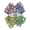
| Noncrystallographic symmetry (NCS) | NCS oper: (Code: given / Matrix: (-0.5621, -0.8271),: | Details | THE ASYMMETRIC UNIT CONTAINS TWO SUBUNITS OF THE TETRAMER. COORDINATES FOR OTHER TWO SUBUNITS CAN BE GENERATED USING CRYSTALLOGRAPHIC OPERATOR (76.02-X; -Y; Z). THE TRANSFORMATION PRESENTED ON *MTRIX* RECORDS BELOW WILL YIELD APPROXIMATE COORDINATES FOR CHAIN *B* WHEN APPLIED TO CHAIN *A* | |
-Components
| #1: Protein | Mass: 51508.754 Da / Num. of mol.: 2 Source method: isolated from a genetically manipulated source Source: (gene. exp.) Citrobacter intermedius (bacteria)References: UniProt: P31012, UniProt: P31013*PLUS, tyrosine phenol-lyase#2: Chemical | ChemComp-SO4 / Sulfate  Mass: 96.063 Da / Num. of mol.: 4 / Source method: obtained synthetically / Formula: SO4 Mass: 96.063 Da / Num. of mol.: 4 / Source method: obtained synthetically / Formula: SO4#3: Water | ChemComp-HOH / | Water Mass: 18.015 Da / Num. of mol.: 450 / Source method: isolated from a natural source / Formula: H2O Mass: 18.015 Da / Num. of mol.: 450 / Source method: isolated from a natural source / Formula: H2O |
|---|
-Experimental details
-Experiment
| Experiment | Method: X-RAY DIFFRACTION |
|---|
- Sample preparation
| Crystal | Density Matthews: 2.39 Å3/Da / Density % sol: 48.44 % | ||||||||||||||||||||||||||||||
|---|---|---|---|---|---|---|---|---|---|---|---|---|---|---|---|---|---|---|---|---|---|---|---|---|---|---|---|---|---|---|---|
| Crystal grow | *PLUS pH: 7 / Method: vapor diffusion, sitting drop | ||||||||||||||||||||||||||||||
| Components of the solutions | *PLUS
|
-Data collection
| Reflection | *PLUS Highest resolution: 2.3 Å / Lowest resolution: 15 Å / Redundancy: 4.1 % / Rmerge(I) obs: 0.058 |
|---|
- Processing
| Software | Name: PROLSQ / Classification: refinement | |||||||||||||||||||||||||||||||||||||||||||||||||||||||||||||||
|---|---|---|---|---|---|---|---|---|---|---|---|---|---|---|---|---|---|---|---|---|---|---|---|---|---|---|---|---|---|---|---|---|---|---|---|---|---|---|---|---|---|---|---|---|---|---|---|---|---|---|---|---|---|---|---|---|---|---|---|---|---|---|---|---|
| Refinement | Resolution: 2.3→10 Å Details: ABOUT 6% OF THE AMINO ACIDS HAVE POOR ELECTRON DENSITY AND COULD NOT BE LOCATED. THESE RESIDUES LIE IN THREE LOOPS ON THE SURFACE OF THE MOLECULE: RESIDUES 123 - 131, 384 - 398, 442 - 447 IN ...Details: ABOUT 6% OF THE AMINO ACIDS HAVE POOR ELECTRON DENSITY AND COULD NOT BE LOCATED. THESE RESIDUES LIE IN THREE LOOPS ON THE SURFACE OF THE MOLECULE: RESIDUES 123 - 131, 384 - 398, 442 - 447 IN THE A CHAIN AND RESIDUES 123 - 133, 384 - 398, 442 - 445 IN THE B CHAIN. NO COORDINATES ARE PRESENT FOR THESE RESIDUES.
| |||||||||||||||||||||||||||||||||||||||||||||||||||||||||||||||
| Refinement step | Cycle: LAST / Resolution: 2.3→10 Å
| |||||||||||||||||||||||||||||||||||||||||||||||||||||||||||||||
| Refine LS restraints |
| |||||||||||||||||||||||||||||||||||||||||||||||||||||||||||||||
| Refinement | *PLUS Highest resolution: 2.3 Å / Lowest resolution: 10 Å / Num. reflection obs: 43205 / Rfactor obs: 0.1622 | |||||||||||||||||||||||||||||||||||||||||||||||||||||||||||||||
| Solvent computation | *PLUS | |||||||||||||||||||||||||||||||||||||||||||||||||||||||||||||||
| Displacement parameters | *PLUS | |||||||||||||||||||||||||||||||||||||||||||||||||||||||||||||||
| Refine LS restraints | *PLUS
|
