 Movie
Movie Controller

[English] 日本語
 Yorodumi
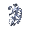
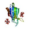
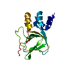
- PDB-1tol: FUSION OF N-TERMINAL DOMAIN OF THE MINOR COAT PROTEIN FROM GENE I... -
+ Open data

- Basic information
| Entry | Database: PDB / ID: 1tol | ||||||
|---|---|---|---|---|---|---|---|
| Title | FUSION OF N-TERMINAL DOMAIN OF THE MINOR COAT PROTEIN FROM GENE III IN PHAGE M13, AND C-TERMINAL DOMAIN OF E. COLI PROTEIN-TOLA | ||||||
 Components Components | PROTEIN (FUSION PROTEIN CONSISTING OF MINOR COAT PROTEIN, GLYCINE RICH LINKER, TOLA, AND A HIS TAG) | ||||||
 Keywords Keywords |  VIRAL PROTEIN / BACTERIOPHAGE M13 / PHAGE INFECTION / TOL PATHWAY / FUSION PROTEIN VIRAL PROTEIN / BACTERIOPHAGE M13 / PHAGE INFECTION / TOL PATHWAY / FUSION PROTEIN | ||||||
| Function / homology |  Function and homology information Function and homology informationcellular response to bacteriocin / regulation of membrane invagination / bacteriocin transport / toxin transmembrane transporter activity / protein import / virion binding / cell division site / : / viral capsid / disordered domain specific binding ...cellular response to bacteriocin / regulation of membrane invagination / bacteriocin transport / toxin transmembrane transporter activity / protein import / virion binding / cell division site / : / viral capsid / disordered domain specific binding / cell cycle / symbiont entry into host cell / cell division / protein domain specific binding / membrane / plasma membraneSimilarity search - Function | ||||||
| Biological species |  Enterobacteria phage M13 (virus) Enterobacteria phage M13 (virus) Escherichia coli (E. coli) Escherichia coli (E. coli) | ||||||
| Method | X-RAY DIFFRACTION / SYNCHROTRON / MIRAS / Resolution: 1.85 Å | ||||||
 Authors Authors | Lubkowski, J. / Wlodawer, A. / Hennecke, F. / Plueckthun, A. | ||||||
 Citation Citation | Journal: Structure Fold.Des. / Year: 1999 Title: Filamentous phage infection: crystal structure of g3p in complex with its coreceptor, the C-terminal domain of TolA. Authors: Lubkowski, J. / Hennecke, F. / Pluckthun, A. / Wlodawer, A. | ||||||
| History |
|
- Structure visualization
| Structure viewer | Molecule: MolmilJmol/JSmol |
|---|
- Downloads & links
-Download
| PDBx/mmCIF format | 1tol.cif.gz | 47 KB | Display | PDBx/mmCIF format |
|---|---|---|---|---|
| PDB format | pdb1tol.ent.gz | 32.5 KB | Display | PDB format |
| PDBx/mmJSON format | 1tol.json.gz | Tree view | PDBx/mmJSON format | |
| Others |  Other downloads Other downloads |
-Validation report
| Arichive directory | https://data.pdbj.org/pub/pdb/validation_reports/to/1tolftp://data.pdbj.org/pub/pdb/validation_reports/to/1tol | HTTPS FTP |
|---|
-Related structure data
| Similar structure data |
|---|


-Links
 PDBj
PDBj- Assembly
| Deposited unit | 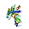
| ||||||||
|---|---|---|---|---|---|---|---|---|---|
| 1 |
| ||||||||
| Unit cell |
|
-Components
| #1: Protein | Mass: 22851.123 Da / Num. of mol.: 1 Fragment: N-TERMINAL DOMAIN OF MINOR COAT PROTEIN AND C-TERMINAL DOMAIN OF TOLA Source method: isolated from a genetically manipulated source Details: FUSION PROTEIN COMPRISES RESIDUES 1-86 OF MATURE MINOR COAT PROTEIN FROM GENE III, INCLUDING GLYCINE-RICH LINKER (GGGSEGGGSEGGGSEGGG), AND RESIDUES 295-421 OF TOLA, AND C-TERMINAL TAIL WITH SEQUENCE AAAHHHHHH Source: (gene. exp.) Enterobacteria phage M13 (virus), (gene. exp.) Escherichia coli (E. coli)Genus: Inovirus, Escherichia Ff phages / Species: , / Description: FUSION GENE / Plasmid: PTFT74 / Production host: Escherichia coli (E. coli) / Strain (production host): BL21(DE3) / References: UniProt: A0A0N8P2C2, UniProt: P19934 |
|---|---|
| #2: Water | ChemComp-HOH / Water Mass: 18.015 Da / Num. of mol.: 159 / Source method: isolated from a natural source / Formula: H2O Mass: 18.015 Da / Num. of mol.: 159 / Source method: isolated from a natural source / Formula: H2O |
-Experimental details
-Experiment
| Experiment | Method: X-RAY DIFFRACTION / Number of used crystals: 1 |
|---|
- Sample preparation
| Crystal | Density Matthews: 3.22 Å3/Da / Density % sol: 61.8 % Description: MIRAS WAS SUPPORTED BY MOLECULAR REPLACEMENT USING N-TERMINAL DOMAIN FROM PREVIOUSLY SOLVED STRUCTURE OF G3P (1G3P) AS A STARTING MODEL |
|---|---|
| Crystal grow | pH: 7.5 Details: PROTEIN AT CONCENTRATION 8-12 MG/ML IN 50 MM HEPES (PH=7.5) WITH ADDITION OF DTT C=2MM, WAS CRYSTALLIZED USING VAPOR DIFFUSION FROM THE SITTING OR HANGING DROP, WITH THE SOLUTION CONTAINING ...Details: PROTEIN AT CONCENTRATION 8-12 MG/ML IN 50 MM HEPES (PH=7.5) WITH ADDITION OF DTT C=2MM, WAS CRYSTALLIZED USING VAPOR DIFFUSION FROM THE SITTING OR HANGING DROP, WITH THE SOLUTION CONTAINING 25% PEG4000, 0.08M TRIS (PH=8.5), AND 0.15 M SODIUM ACETATE AS A PRECIPITANT. BEFORE SETTING THE DROPS, PROTEIN SOLUTION WAS MIXED WITH THE PRECIPITANT IN THE RATIO 1:1. CRYSTALS WERE GROWING BEST AT TEMPERATURE 15 DEGREES CELSIUS. |
-Data collection
| Diffraction | Mean temperature: 100 K |
|---|---|
| Diffraction source | Source: SYNCHROTRON / Site: CHESS  / Beamline: F2 / Wavelength: 0.98 / Beamline: F2 / Wavelength: 0.98 |
| Detector | Type: ADSC / Detector: CCD / Date: Apr 1, 1998 / Details: MIRROR |
| Radiation | Monochromator: SI CRYSTAL / Protocol: SINGLE WAVELENGTH / Monochromatic (M) / Laue (L): M / Scattering type: x-ray |
| Radiation wavelength | Wavelength: 0.98 Å / Relative weight: 1 |
| Reflection | Resolution: 1.85→25 Å / Num. obs: 22243 / % possible obs: 99.6 % / Observed criterion σ(I): -3 / Redundancy: 15.1 % / Rmerge(I) obs: 0.042 / Net I/σ(I): 36.4 |
| Reflection shell | Resolution: 1.85→1.92 Å / Redundancy: 8.05 % / Rmerge(I) obs: 0.446 / Mean I/σ(I) obs: 5.85 / % possible all: 100 |
| Reflection | *PLUS Num. measured all: 190164 |
| Reflection shell | *PLUS % possible obs: 100 % |
- Processing
| Software |
| |||||||||||||||||||||||||||||||||
|---|---|---|---|---|---|---|---|---|---|---|---|---|---|---|---|---|---|---|---|---|---|---|---|---|---|---|---|---|---|---|---|---|---|---|
| Refinement | Method to determine structure: MIRAS / Resolution: 1.85→10 Å / Num. parameters: 5335 / Num. restraintsaints: 4829 / Cross valid method: FREE-R / σ(F): 0 / Stereochemistry target values: ENGH AND HUBER Details: ANISOTROPIC SCALING APPLIED BY THE METHOD OF PARKIN, MOEZZI & HOPE, J.APPL.CRYST.28 (1995)53-56
| |||||||||||||||||||||||||||||||||
| Solvent computation | Solvent model: MOEWS & KRETSINGER, J.MOL.BIOL.91(1973)201-228 | |||||||||||||||||||||||||||||||||
| Refine analyze | Num. disordered residues: 0 / Occupancy sum hydrogen: 0 / Occupancy sum non hydrogen: 1325 | |||||||||||||||||||||||||||||||||
| Refinement step | Cycle: LAST / Resolution: 1.85→10 Å
| |||||||||||||||||||||||||||||||||
| Refine LS restraints |
|
