 Movie
Movie Controller

[English] 日本語
 Yorodumi
- PDB-1tmi: Structure of Thermotoga maritima S63A non-processing mutant S-ade... -
+ Open data

- Basic information
| Entry | Database: PDB / ID: 1tmi | ||||||
|---|---|---|---|---|---|---|---|
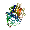
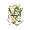
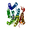
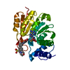
| Title | Structure of Thermotoga maritima S63A non-processing mutant S-adenosylmethionine decarboxylase | ||||||
 Components Components | S-adenosylmethionine decarboxylase proenzyme, AdoMetDC, SamDC | ||||||
 Keywords Keywords |  LYASE / two-layer alpha beta-sandwich LYASE / two-layer alpha beta-sandwich | ||||||
| Function / homology |  Function and homology informationadenosylmethionine decarboxylase / adenosylmethionine decarboxylase activity / spermidine biosynthetic process / cytosol Function and homology informationadenosylmethionine decarboxylase / adenosylmethionine decarboxylase activity / spermidine biosynthetic process / cytosolSimilarity search - Function | ||||||
| Biological species |   Thermotoga maritima (bacteria) Thermotoga maritima (bacteria) | ||||||
| Method | X-RAY DIFFRACTION / SYNCHROTRON / MAD / Resolution: 1.7 Å | ||||||
 Authors Authors | Toms, A.V. / Kinsland, C. / McCloskey, D.E. / Pegg, A.E. / Ealick, S.E. | ||||||
 Citation Citation | Journal: J.Biol.Chem. / Year: 2004 Title: Evolutionary Links as Revealed by the Structure of Thermotoga maritima S-Adenosylmethionine Decarboxylase. Authors: Toms, A.V. / Kinsland, C. / McCloskey, D.E. / Pegg, A.E. / Ealick, S.E. | ||||||
| History |
|
- Structure visualization
| Structure viewer | Molecule: MolmilJmol/JSmol |
|---|
- Downloads & links
-Download
| PDBx/mmCIF format | 1tmi.cif.gz | 61.9 KB | Display | PDBx/mmCIF format |
|---|---|---|---|---|
| PDB format | pdb1tmi.ent.gz | 46.6 KB | Display | PDB format |
| PDBx/mmJSON format | 1tmi.json.gz | Tree view | PDBx/mmJSON format | |
| Others |  Other downloads Other downloads |
-Validation report
| Arichive directory | https://data.pdbj.org/pub/pdb/validation_reports/tm/1tmiftp://data.pdbj.org/pub/pdb/validation_reports/tm/1tmi | HTTPS FTP |
|---|
-Related structure data


-Links
 PDBj
PDBj- Assembly
| Deposited unit | 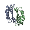
| ||||||||
|---|---|---|---|---|---|---|---|---|---|
| 1 |
| ||||||||
| Unit cell |
| ||||||||
| Details | The biological assembly is a dimer. The asymmetric unit contains 1 biological unit. |
-Components
| #1: Protein | Mass: 14790.736 Da / Num. of mol.: 2 / Mutation: S63A Source method: isolated from a genetically manipulated source Source: (gene. exp.) Thermotoga maritima (bacteria) / Gene: SPEH, TM0655 / Plasmid: pET-28a / Production host: Escherichia coli (E. coli) / Strain (production host): BL21Star(DE3)pRareReferences: UniProt: Q9WZC3, adenosylmethionine decarboxylase#2: Water | ChemComp-HOH / | Water Mass: 18.015 Da / Num. of mol.: 148 / Source method: isolated from a natural source / Formula: H2O Mass: 18.015 Da / Num. of mol.: 148 / Source method: isolated from a natural source / Formula: H2O |
|---|
-Experimental details
-Experiment
| Experiment | Method: X-RAY DIFFRACTION / Number of used crystals: 1 |
|---|
- Sample preparation
| Crystal | Density Matthews: 2.49 Å3/Da / Density % sol: 51 % |
|---|---|
| Crystal grow | Temperature: 295.4 K / Method: vapor diffusion, hanging drop / pH: 8 Details: ammonium formate, HEPES, pH 8, VAPOR DIFFUSION, HANGING DROP, temperature 295.4K |
-Data collection
| Diffraction | Mean temperature: 110 K | ||||||||||||
|---|---|---|---|---|---|---|---|---|---|---|---|---|---|
| Diffraction source | Source: SYNCHROTRON / Site: APS  / Beamline: 8-BM / Wavelength: 0.9792,0.9793,0.9641 / Beamline: 8-BM / Wavelength: 0.9792,0.9793,0.9641 | ||||||||||||
| Detector | Type: ADSC QUANTUM 315 / Detector: CCD / Date: Aug 22, 2003 | ||||||||||||
| Radiation | Protocol: MAD / Monochromatic (M) / Laue (L): M / Scattering type: x-ray | ||||||||||||
| Radiation wavelength |
| ||||||||||||
| Reflection | Resolution: 1.7→32.5 Å / Num. all: 31186 / Num. obs: 30984 / % possible obs: 98.7 % / Observed criterion σ(F): 1 / Observed criterion σ(I): 1 / Redundancy: 5.7 % / Biso Wilson estimate: 22.6 Å2 / Rsym value: 0.048 / Net I/σ(I): 30.7 | ||||||||||||
| Reflection shell | Resolution: 1.7→1.81 Å / Redundancy: 5.6 % / Mean I/σ(I) obs: 4.7 / Rsym value: 0.385 / % possible all: 100 |
- Processing
| Software |
| ||||||||||||||||||||||||||||||||||||
|---|---|---|---|---|---|---|---|---|---|---|---|---|---|---|---|---|---|---|---|---|---|---|---|---|---|---|---|---|---|---|---|---|---|---|---|---|---|
| Refinement | Method to determine structure: MAD / Resolution: 1.7→32.47 Å / Rfactor Rfree error: 0.005 / Data cutoff high absF: 410997.27 / Data cutoff low absF: 0 / Isotropic thermal model: anisotropic / Cross valid method: THROUGHOUT / σ(F): 0 / Stereochemistry target values: Engh & Huber Details: twinning operator was used to ensure that pairs of twin related reflections were in the same set
| ||||||||||||||||||||||||||||||||||||
| Solvent computation | Solvent model: FLAT MODEL / Bsol: 62.8139 Å2 / ksol: 0.391508 e/Å3 | ||||||||||||||||||||||||||||||||||||
| Displacement parameters | Biso mean: 28.5 Å2
| ||||||||||||||||||||||||||||||||||||
| Refine analyze |
| ||||||||||||||||||||||||||||||||||||
| Refinement step | Cycle: LAST / Resolution: 1.7→32.47 Å
| ||||||||||||||||||||||||||||||||||||
| Refine LS restraints |
| ||||||||||||||||||||||||||||||||||||
| LS refinement shell | Resolution: 1.7→1.81 Å / Rfactor Rfree error: 0.014 / Total num. of bins used: 6
| ||||||||||||||||||||||||||||||||||||
| Xplor file |
|
