 Movie
Movie Controller

[English] 日本語
 Yorodumi
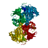
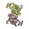
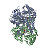
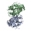
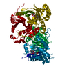
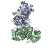
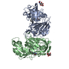
- PDB-1tg5: Crystal structures of plant 4-hydroxyphenylpyruvate dioxygenases ... -
+ Open data

- Basic information
| Entry | Database: PDB / ID: 1tg5 | ||||||
|---|---|---|---|---|---|---|---|
| Title | Crystal structures of plant 4-hydroxyphenylpyruvate dioxygenases complexed with DAS645 | ||||||
 Components Components | 4-hydroxyphenylpyruvate dioxygenase | ||||||
 Keywords Keywords | OXIDOREDUCTASE / Arabidopsis thaliana / 4-hydroxyphenylpyruvate dioxygenase / HPPD / atHPPD | ||||||
| Function / homology |  Function and homology information4-hydroxyphenylpyruvate dioxygenase / 4-hydroxyphenylpyruvate dioxygenase activity / tyrosine catabolic process / L-phenylalanine catabolic process / iron ion binding / identical protein binding / cytoplasm Function and homology information4-hydroxyphenylpyruvate dioxygenase / 4-hydroxyphenylpyruvate dioxygenase activity / tyrosine catabolic process / L-phenylalanine catabolic process / iron ion binding / identical protein binding / cytoplasmSimilarity search - Function | ||||||
| Biological species |  Arabidopsis thaliana (thale cress) Arabidopsis thaliana (thale cress) | ||||||
| Method | X-RAY DIFFRACTION / MOLECULAR REPLACEMENT / Resolution: 1.9 Å | ||||||
 Authors Authors | Yang, C. / Pflugrath, J.W. / Camper, D.L. / Foster, M.L. / Pernich, D.J. / Walsh, T.A. | ||||||
 Citation Citation | Journal: Biochemistry / Year: 2004 Title: Structural basis for herbicidal inhibitor selectivity revealed by comparison of crystal structures of plant and Mammalian 4-hydroxyphenylpyruvate dioxygenases Authors: Yang, C. / Pflugrath, J.W. / Camper, D.L. / Foster, M.L. / Pernich, D.J. / Walsh, T.A. | ||||||
| History |
|
- Structure visualization
| Structure viewer | Molecule: MolmilJmol/JSmol |
|---|
- Downloads & links
-Download
| PDBx/mmCIF format | 1tg5.cif.gz | 91.4 KB | Display | PDBx/mmCIF format |
|---|---|---|---|---|
| PDB format | pdb1tg5.ent.gz | 67.9 KB | Display | PDB format |
| PDBx/mmJSON format | 1tg5.json.gz | Tree view | PDBx/mmJSON format | |
| Others |  Other downloads Other downloads |
-Validation report
| Arichive directory | https://data.pdbj.org/pub/pdb/validation_reports/tg/1tg5ftp://data.pdbj.org/pub/pdb/validation_reports/tg/1tg5 | HTTPS FTP |
|---|
-Related structure data
| Related structure data |  1sqdSC  1sqiC 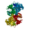 1tfzC S: Starting model for refinement C: citing same article ( |
|---|---|
| Similar structure data |


-Links
 PDBj
PDBj


- Assembly
| Deposited unit | 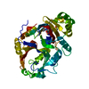
| ||||||||
|---|---|---|---|---|---|---|---|---|---|
| 1 | 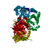
| ||||||||
| Unit cell |
| ||||||||
| Details | Homodimer. The second part of the biological assembly is generated by the two fold axis. |
-Components
| #1: Protein | / 4HPPD / HPD / HPPDase Mass: 46783.797 Da / Num. of mol.: 1 Source method: isolated from a genetically manipulated source Source: (gene. exp.) Arabidopsis thaliana (thale cress) / Gene: HPD, AT1G06570, F12K11.9 / Plasmid: pET28 / Species (production host): Escherichia coli / Production host:  Escherichia coli BL21(DE3) (bacteria) / Strain (production host): BL21DE3 Escherichia coli BL21(DE3) (bacteria) / Strain (production host): BL21DE3References: UniProt: P93836, 4-hydroxyphenylpyruvate dioxygenase |
|---|---|
| #2: Chemical | ChemComp-FE2 /   Mass: 55.845 Da / Num. of mol.: 1 / Source method: obtained synthetically / Formula: Fe Mass: 55.845 Da / Num. of mol.: 1 / Source method: obtained synthetically / Formula: Fe |
| #3: Chemical | ChemComp-645 / [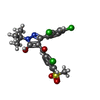  Mass: 501.811 Da / Num. of mol.: 1 / Source method: obtained synthetically / Formula: C21H19Cl3N2O4S Mass: 501.811 Da / Num. of mol.: 1 / Source method: obtained synthetically / Formula: C21H19Cl3N2O4S |
| #4: Water | ChemComp-HOH / Water Mass: 18.015 Da / Num. of mol.: 260 / Source method: isolated from a natural source / Formula: H2O Mass: 18.015 Da / Num. of mol.: 260 / Source method: isolated from a natural source / Formula: H2O |
-Experimental details
-Experiment
| Experiment | Method: X-RAY DIFFRACTION / Number of used crystals: 1 |
|---|
- Sample preparation
| Crystal | Density Matthews: 2.3 Å3/Da / Density % sol: 40.5 % |
|---|---|
| Crystal grow | Temperature: 298 K / Method: vapor diffusion, hanging drop / pH: 7.3 Details: PEG 4000, isopropanol, Bis-tris, potassium chloride, cobalt chloride, pH 7.3, VAPOR DIFFUSION, HANGING DROP, temperature 298K |
-Data collection
| Diffraction | Mean temperature: 100 K |
|---|---|
| Diffraction source | Source: ROTATING ANODE / Type: RIGAKU RU200 / Wavelength: 1.5418 Å |
| Detector | Type: RIGAKU RAXIS IV / Detector: IMAGE PLATE / Date: Jan 1, 2002 / Details: multilayer optics |
| Radiation | Monochromator: Multilayer optics / Protocol: SINGLE WAVELENGTH / Monochromatic (M) / Laue (L): M / Scattering type: x-ray |
| Radiation wavelength | Wavelength: 1.5418 Å / Relative weight: 1 |
| Reflection | Resolution: 1.9→15 Å / Num. all: 30430 / Num. obs: 30159 / % possible obs: 98.3 % / Observed criterion σ(F): 2 / Observed criterion σ(I): 2 / Redundancy: 4.4 % / Rmerge(I) obs: 0.048 / Net I/σ(I): 12 |
| Reflection shell | Resolution: 1.9→1.95 Å / Redundancy: 2 % / Rmerge(I) obs: 0.155 / Mean I/σ(I) obs: 2.5 / Num. unique all: 1923 / % possible all: 83.8 |
- Processing
| Software |
| |||||||||||||||||||||
|---|---|---|---|---|---|---|---|---|---|---|---|---|---|---|---|---|---|---|---|---|---|---|
| Refinement | Method to determine structure: MOLECULAR REPLACEMENT Starting model: PDB ENTRY 1SQD Resolution: 1.9→15 Å / Cross valid method: THROUGHOUT / σ(F): 2 / Stereochemistry target values: Engh & Huber
| |||||||||||||||||||||
| Refinement step | Cycle: LAST / Resolution: 1.9→15 Å
|
