 Movie
Movie Controller


+ Open data

- Basic information
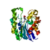
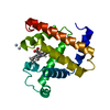
| Entry | Database: PDB / ID: 1td5 | ||||||
|---|---|---|---|---|---|---|---|
| Title | Crystal Structure of the Ligand Binding Domain of E. coli IclR. | ||||||
 Components Components | Acetate operon repressor | ||||||
 Keywords Keywords |  TRANSCRIPTION / MIDWEST CENTER FOR STRUCTURAL GENOMICS / ALPHA/BETA DOMAIN / LIGAND BINDING DOMAIN / TRANSCRIPTION REGULATOR / PSI / Protein Structure Initiative / MCSG TRANSCRIPTION / MIDWEST CENTER FOR STRUCTURAL GENOMICS / ALPHA/BETA DOMAIN / LIGAND BINDING DOMAIN / TRANSCRIPTION REGULATOR / PSI / Protein Structure Initiative / MCSG | ||||||
| Function / homology |  Function and homology informationglyoxylate cycle / DNA-binding transcription factor activity / negative regulation of DNA-templated transcription / regulation of DNA-templated transcription / DNA binding / cytosol Function and homology informationglyoxylate cycle / DNA-binding transcription factor activity / negative regulation of DNA-templated transcription / regulation of DNA-templated transcription / DNA binding / cytosolSimilarity search - Function | ||||||
| Biological species |  Escherichia coli (E. coli) Escherichia coli (E. coli) | ||||||
| Method | X-RAY DIFFRACTION / SYNCHROTRON / MAD / Resolution: 2.3 Å | ||||||
 Authors Authors | Walker, J.R. / Evdokimova, L. / Zhang, R.-G. / Bochkarev, A. / Joachimiak, A. / Arrowsmith, C. / Edwards, A. / Savchenko, A. / Midwest Center for Structural Genomics (MCSG) | ||||||
 Citation Citation | Journal: To be Published Title: Structural Analyses of the Ligand Binding Sites of the IclR family of transcriptional regulators Authors: Walker, J.R. / Evdokimova, L. / Zhang, R.-G. / Bochkarev, A. / Arrowsmith, C. / Edwards, A. / Savchenko, A. #1: Journal: J.Biol.Chem. / Year: 2002Title: Crystal Structure of Thermotoga maritima 0065, a member of the IclR transcriptional factor family Authors: Zhang, R.-G. / Youngchang, K. / Skarina, T. / Beasley, S. / Laskowski, R. / Arrowsmith, C. / Edwards, A. / Joachimiak, A. / Savchenko, A. #2: Journal: To be PublishedTitle: Crystal Structure Analysis of the E. coli Glyoxylate Regulatory Protein Ligand Binding Domain Authors: Walker, J.R. / Skarina, T. / Kudrytska, M. / Arrowsmith, C. / Edwards, A. / Savchenko, A. | ||||||
| History |
| ||||||
| Remark 300 | BIOMOLECULE: 1, 2, 3, 4 THIS ENTRY CONTAINS THE CRYSTALLOGRAPHIC ASYMMETRIC UNIT WHICH CONSISTS OF ...BIOMOLECULE: 1, 2, 3, 4 THIS ENTRY CONTAINS THE CRYSTALLOGRAPHIC ASYMMETRIC UNIT WHICH CONSISTS OF 4 CHAIN(S). SEE REMARK 350 FOR INFORMATION ON GENERATING THE BIOLOGICAL MOLECULE(S). GEL FILTRATION STUDIES SHOW THAT THE LIGAND BINDING DOMAIN OF ICLR IS A MONOMER IN SOLUTION. |
- Structure visualization
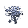
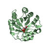
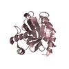
| Structure viewer | Molecule: MolmilJmol/JSmol |
|---|
- Downloads & links
-Download
| PDBx/mmCIF format | 1td5.cif.gz | 151.6 KB | Display | PDBx/mmCIF format |
|---|---|---|---|---|
| PDB format | pdb1td5.ent.gz | 125.9 KB | Display | PDB format |
| PDBx/mmJSON format | 1td5.json.gz | Tree view | PDBx/mmJSON format | |
| Others |  Other downloads Other downloads |
-Validation report
| Arichive directory | https://data.pdbj.org/pub/pdb/validation_reports/td/1td5ftp://data.pdbj.org/pub/pdb/validation_reports/td/1td5 | HTTPS FTP |
|---|
-Related structure data
| Related structure data |  1t9l |
|---|---|
| Similar structure data | |
| Other databases |


-Links
 PDBj
PDBj

- Assembly
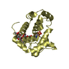
| Deposited unit | 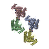
| ||||||||
|---|---|---|---|---|---|---|---|---|---|
| 1 | 
| ||||||||
| 2 | 
| ||||||||
| 3 | 
| ||||||||
| 4 | 
| ||||||||
| Unit cell |
|
-Components
| #1: Protein | Mass: 22716.697 Da / Num. of mol.: 4 / Fragment: ligand binding domain Source method: isolated from a genetically manipulated source Source: (gene. exp.) Escherichia coli (E. coli) / Gene: ICLR, B4018 / Species (production host): Escherichia coli / Production host: Escherichia coli BL21(DE3) (bacteria) / Strain (production host): BL21(DE3) / References: UniProt: P16528#2: Water | ChemComp-HOH / | Water Mass: 18.015 Da / Num. of mol.: 335 / Source method: isolated from a natural source / Formula: H2O Mass: 18.015 Da / Num. of mol.: 335 / Source method: isolated from a natural source / Formula: H2O |
|---|
-Experimental details
-Experiment
| Experiment | Method: X-RAY DIFFRACTION / Number of used crystals: 1 |
|---|
- Sample preparation
| Crystal | Density Matthews: 2.3 Å3/Da / Density % sol: 46.6 % |
|---|---|
| Crystal grow | Temperature: 298 K / Method: vapor diffusion, hanging drop / pH: 7.5 Details: HEPES pH 7.5, Potassium acetate, PEG 3350, VAPOR DIFFUSION, HANGING DROP, temperature 298.0K |
-Data collection
| Diffraction | Mean temperature: 100 K | ||||||||||||
|---|---|---|---|---|---|---|---|---|---|---|---|---|---|
| Diffraction source | Source: SYNCHROTRON / Site: APS  / Beamline: 19-ID / Wavelength: 0.97934,0.9791,0.96110 / Beamline: 19-ID / Wavelength: 0.97934,0.9791,0.96110 | ||||||||||||
| Detector | Type: MARRESEARCH / Detector: CCD / Date: Jul 4, 2002 / Details: mirrors | ||||||||||||
| Radiation | Monochromator: SAGITALLY FOCUSED Si(111) / Protocol: MAD / Monochromatic (M) / Laue (L): M / Scattering type: x-ray | ||||||||||||
| Radiation wavelength |
| ||||||||||||
| Reflection | Resolution: 2.3→20 Å / Num. all: 31820 / Num. obs: 31820 / % possible obs: 99.2 % / Observed criterion σ(F): 0 / Observed criterion σ(I): -3 / Redundancy: 8.96 % / Biso Wilson estimate: 21.5 Å2 / Rmerge(I) obs: 0.092 / Net I/σ(I): 19.9 | ||||||||||||
| Reflection shell | Resolution: 2.3→2.38 Å / Rmerge(I) obs: 0.38 / Mean I/σ(I) obs: 4 / Num. unique all: 3100 / % possible all: 98.5 |
- Processing
| Software |
| ||||||||||||||||||||||||||||||||||||
|---|---|---|---|---|---|---|---|---|---|---|---|---|---|---|---|---|---|---|---|---|---|---|---|---|---|---|---|---|---|---|---|---|---|---|---|---|---|
| Refinement | Method to determine structure: MAD / Resolution: 2.3→19.91 Å / Rfactor Rfree error: 0.009 / Data cutoff high absF: 187632.18 / Data cutoff low absF: 0 / Isotropic thermal model: RESTRAINED / Cross valid method: THROUGHOUT / σ(F): 0 / Stereochemistry target values: Engh & Huber
| ||||||||||||||||||||||||||||||||||||
| Solvent computation | Solvent model: FLAT MODEL / Bsol: 47.2132 Å2 / ksol: 0.332517 e/Å3 | ||||||||||||||||||||||||||||||||||||
| Displacement parameters | Biso mean: 37.7 Å2
| ||||||||||||||||||||||||||||||||||||
| Refine analyze |
| ||||||||||||||||||||||||||||||||||||
| Refinement step | Cycle: LAST / Resolution: 2.3→19.91 Å
| ||||||||||||||||||||||||||||||||||||
| Refine LS restraints |
| ||||||||||||||||||||||||||||||||||||
| LS refinement shell | Resolution: 2.3→2.44 Å / Rfactor Rfree error: 0.031 / Total num. of bins used: 6
| ||||||||||||||||||||||||||||||||||||
| Xplor file |
|
