 Movie
Movie Controller

[English] 日本語
 Yorodumi
- PDB-1t13: Crystal Structure Of Lumazine Synthase From Brucella Abortus Boun... -
+ Open data

- Basic information
| Entry | Database: PDB / ID: 1t13 | ||||||
|---|---|---|---|---|---|---|---|
| Title | Crystal Structure Of Lumazine Synthase From Brucella Abortus Bound To 5-nitro-6-(D-ribitylamino)-2,4(1H,3H) pyrimidinedione | ||||||
 Components Components | 6,7-dimethyl-8-ribityllumazine synthase Lumazine synthase Lumazine synthase | ||||||
 Keywords Keywords | TRANSFERASE | ||||||
| Function / homology |  Function and homology information6,7-dimethyl-8-ribityllumazine synthase / 6,7-dimethyl-8-ribityllumazine synthase activity / riboflavin synthase complex / riboflavin biosynthetic process / cytosol Function and homology information6,7-dimethyl-8-ribityllumazine synthase / 6,7-dimethyl-8-ribityllumazine synthase activity / riboflavin synthase complex / riboflavin biosynthetic process / cytosolSimilarity search - Function | ||||||
| Biological species |  Brucella abortus (bacteria) Brucella abortus (bacteria) | ||||||
| Method | X-RAY DIFFRACTION / SYNCHROTRON / MOLECULAR REPLACEMENT / Resolution: 2.9 Å | ||||||
 Authors Authors | Klinke, S. / Zylberman, V. / Vega, D.R. / Guimaraes, B.G. / Braden, B.C. / Goldbaum, F.A. | ||||||
 Citation Citation | Journal: J.Mol.Biol. / Year: 2005 Title: Crystallographic studies on Decameric Brucella spp. Lumazine Synthase: A Novel Quaternary Arrangement Evolved for a New Function? Authors: Klinke, S. / Zylberman, V. / Vega, D.R. / Guimaraes, B.G. / Braden, B.C. / Goldbaum, F.A. | ||||||
| History |
|
- Structure visualization
| Structure viewer | Molecule: MolmilJmol/JSmol |
|---|
- Downloads & links
-Download
| PDBx/mmCIF format | 1t13.cif.gz | 153.7 KB | Display | PDBx/mmCIF format |
|---|---|---|---|---|
| PDB format | pdb1t13.ent.gz | 123.8 KB | Display | PDB format |
| PDBx/mmJSON format | 1t13.json.gz | Tree view | PDBx/mmJSON format | |
| Others |  Other downloads Other downloads |
-Validation report
| Arichive directory | https://data.pdbj.org/pub/pdb/validation_reports/t1/1t13ftp://data.pdbj.org/pub/pdb/validation_reports/t1/1t13 | HTTPS FTP |
|---|
-Related structure data
| Related structure data | 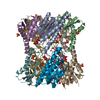 1xn1C 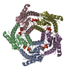 1di0S S: Starting model for refinement C: citing same article ( |
|---|---|
| Similar structure data |


-Links
 PDBj
PDBj- Assembly
| Deposited unit | 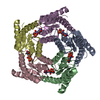
| ||||||||
|---|---|---|---|---|---|---|---|---|---|
| 1 | 
| ||||||||
| Unit cell |
| ||||||||
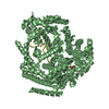
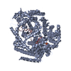
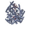
| Details | The biological assembly is a dimer of pentamers which can be generated applying crystallographic symmetry operations to the pentamer in the asymmetric unit. |
-Components
| #1: Protein | Lumazine synthase / DMRL synthase / Lumazine synthase / Riboflavin synthase beta chain Mass: 17381.900 Da / Num. of mol.: 5 Source method: isolated from a genetically manipulated source Source: (gene. exp.) Brucella abortus (bacteria) / Gene: RIBH, BMEII0589, BRA0695 / Plasmid: pET11b / Species (production host): Escherichia coli / Production host: Escherichia coli BL21(DE3) (bacteria) / Strain (production host): BL21(DE3)References: UniProt: Q44668, UniProt: P61711*PLUS, 6,7-dimethyl-8-ribityllumazine synthase#2: Chemical | ChemComp-PO4 / Phosphate  Mass: 94.971 Da / Num. of mol.: 5 / Source method: obtained synthetically / Formula: PO4 Mass: 94.971 Da / Num. of mol.: 5 / Source method: obtained synthetically / Formula: PO4#3: Chemical | ChemComp-INI /   Mass: 306.229 Da / Num. of mol.: 5 / Source method: obtained synthetically / Formula: C9H14N4O8 Mass: 306.229 Da / Num. of mol.: 5 / Source method: obtained synthetically / Formula: C9H14N4O8#4: Water | ChemComp-HOH / | Water Mass: 18.015 Da / Num. of mol.: 89 / Source method: isolated from a natural source / Formula: H2O Mass: 18.015 Da / Num. of mol.: 89 / Source method: isolated from a natural source / Formula: H2O |
|---|
-Experimental details
-Experiment
| Experiment | Method: X-RAY DIFFRACTION / Number of used crystals: 1 |
|---|
- Sample preparation
| Crystal | Density Matthews: 4.2 Å3/Da / Density % sol: 70.8 % |
|---|---|
| Crystal grow | Temperature: 294 K / Method: vapor diffusion, hanging drop / pH: 6.5 Details: 30% PEG 400, 0.1M Na MES pH=6.5, 0.1M Na Acetate, VAPOR DIFFUSION, HANGING DROP, temperature 294K |
-Data collection
| Diffraction | Mean temperature: 115 K |
|---|---|
| Diffraction source | Source: SYNCHROTRON / Site: LNLS  / Beamline: D03B-MX1 / Wavelength: 1.431 Å / Beamline: D03B-MX1 / Wavelength: 1.431 Å |
| Detector | Type: MARRESEARCH / Detector: CCD / Date: Jul 11, 2003 / Details: Si single-crystal |
| Radiation | Protocol: SINGLE WAVELENGTH / Monochromatic (M) / Laue (L): M / Scattering type: x-ray |
| Radiation wavelength | Wavelength: 1.431 Å / Relative weight: 1 |
| Reflection | Resolution: 2.9→52 Å / Num. obs: 34456 / % possible obs: 100 % / Redundancy: 10.7 % / Biso Wilson estimate: 63.4 Å2 / Rmerge(I) obs: 0.093 / Net I/σ(I): 6.6 |
| Reflection shell | Resolution: 2.9→3.06 Å / Redundancy: 10.6 % / Rmerge(I) obs: 0.307 / Mean I/σ(I) obs: 2.4 / Num. unique all: 4980 / % possible all: 100 |
- Processing
| Software |
| |||||||||||||||||||||||||
|---|---|---|---|---|---|---|---|---|---|---|---|---|---|---|---|---|---|---|---|---|---|---|---|---|---|---|
| Refinement | Method to determine structure: MOLECULAR REPLACEMENT Starting model: PDB ENTRY 1DI0 Resolution: 2.9→52 Å / Cross valid method: THROUGHOUT / σ(F): 2 Details: The following residues were not located in the electronic density: Chain A: Met3, Asn4, Gln5, Ser6, Cys7, Pro8, Asn9, Val157. Chain B: Met3, Asn4, Gln5, Ser6, Cys7, Pro8, Asn9, Lys10, Thr11, ...Details: The following residues were not located in the electronic density: Chain A: Met3, Asn4, Gln5, Ser6, Cys7, Pro8, Asn9, Val157. Chain B: Met3, Asn4, Gln5, Ser6, Cys7, Pro8, Asn9, Lys10, Thr11, Leu156, Val157. Chain C: Met3, Asn4, Gln5, Ser6, Cys7, Pro8, Asn9, Lys10, Thr11, Leu156, Val157. Chain D: Met3, Asn4, Gln5, Ser6, Cys7, Pro8, Asn9, Lys10, Leu156, Val157. Chain E: Met3, Asn4, Gln5, Ser6, Cys7, Pro8, Asn9, Lys10, Leu156, Val157. The following residues present poor or missing side-chain electronic density and were modeled as alanine: Chain A: Arg152. Chain B: Glu121. Chain D: Thr11. Chain E: Thr11.
| |||||||||||||||||||||||||
| Displacement parameters | Biso mean: 41.5 Å2 | |||||||||||||||||||||||||
| Refinement step | Cycle: LAST / Resolution: 2.9→52 Å
| |||||||||||||||||||||||||
| Refine LS restraints |
|
