 Movie
Movie Controller


+ Open data

- Basic information
| Entry | Database: PDB / ID: 1szh | ||||||
|---|---|---|---|---|---|---|---|
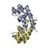
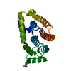
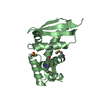
| Title | Crystal Structure of C. elegans HER-1 | ||||||
 Components Components | Her-1 protein | ||||||
 Keywords Keywords |  SIGNALING PROTEIN / extended 3-10 helix / left-handed anti-parallel 4-helix bundle / overhand 3-helix bundle SIGNALING PROTEIN / extended 3-10 helix / left-handed anti-parallel 4-helix bundle / overhand 3-helix bundle | ||||||
| Function / homology |  Function and homology information Function and homology informationmale sex determination / signaling receptor binding / extracellular regionSimilarity search - Function | ||||||
| Biological species |  Caenorhabditis elegans (invertebrata) Caenorhabditis elegans (invertebrata) | ||||||
| Method | X-RAY DIFFRACTION / SYNCHROTRON / MAD / Resolution: 1.5 Å | ||||||
 Authors Authors | Hamaoka, B.Y. / Dann III, C.E. / Geisbrecht, B.V. / Leahy, D.J. | ||||||
 Citation Citation | Journal: Proc.Natl.Acad.Sci.USA / Year: 2004 Title: Crystal structure of Caenorhabditis elegans HER-1 and characterization of the interaction between HER-1 and TRA-2A. Authors: Hamaoka, B.Y. / Dann III, C.E. / Geisbrecht, B.V. / Leahy, D.J. | ||||||
| History |
|
- Structure visualization
| Structure viewer | Molecule: MolmilJmol/JSmol |
|---|
- Downloads & links
-Download
| PDBx/mmCIF format | 1szh.cif.gz | 70.8 KB | Display | PDBx/mmCIF format |
|---|---|---|---|---|
| PDB format | pdb1szh.ent.gz | 57.3 KB | Display | PDB format |
| PDBx/mmJSON format | 1szh.json.gz | Tree view | PDBx/mmJSON format | |
| Others |  Other downloads Other downloads |
-Validation report
| Arichive directory | https://data.pdbj.org/pub/pdb/validation_reports/sz/1szhftp://data.pdbj.org/pub/pdb/validation_reports/sz/1szh | HTTPS FTP |
|---|
-Related structure data
| Similar structure data |
|---|


-Links
 PDBj
PDBj- Assembly
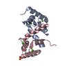
| Deposited unit | 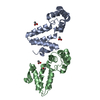
| ||||||||
|---|---|---|---|---|---|---|---|---|---|
| 1 | 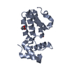
| ||||||||
| 2 | 
| ||||||||
| Unit cell |
|
-Components
| #1: Protein | Mass: 18426.342 Da / Num. of mol.: 2 / Mutation: N98E, N163E Source method: isolated from a genetically manipulated source Source: (gene. exp.) Caenorhabditis elegans (invertebrata) / Gene: HER-1, ZK287.8 / Plasmid: pSGHV0 / Production host:   Cricetulus griseus (Chinese hamster) / References: UniProt: P34704 Cricetulus griseus (Chinese hamster) / References: UniProt: P34704#2: Chemical | ChemComp-ACT / Acetate  Mass: 59.044 Da / Num. of mol.: 4 / Source method: obtained synthetically / Formula: C2H3O2 Mass: 59.044 Da / Num. of mol.: 4 / Source method: obtained synthetically / Formula: C2H3O2#3: Water | ChemComp-HOH / | Water Mass: 18.015 Da / Num. of mol.: 243 / Source method: isolated from a natural source / Formula: H2O Mass: 18.015 Da / Num. of mol.: 243 / Source method: isolated from a natural source / Formula: H2O |
|---|
-Experimental details
-Experiment
| Experiment | Method: X-RAY DIFFRACTION / Number of used crystals: 1 |
|---|
- Sample preparation
| Crystal | Density Matthews: 2.11 Å3/Da / Density % sol: 41.7 % |
|---|---|
| Crystal grow | Temperature: 293 K / Method: vapor diffusion, hanging drop / pH: 4.3 Details: PEG 3350, sodium acetate, ammonijm sulfate, pH 4.3, VAPOR DIFFUSION, HANGING DROP, temperature 293K |
-Data collection
| Diffraction | Mean temperature: 200 K | ||||||||||||
|---|---|---|---|---|---|---|---|---|---|---|---|---|---|
| Diffraction source | Source: SYNCHROTRON / Site: NSLS  / Beamline: X4A / Wavelength: 0.97931, 0.97917, 0.96866 / Beamline: X4A / Wavelength: 0.97931, 0.97917, 0.96866 | ||||||||||||
| Detector | Type: ADSC QUANTUM 4 / Detector: CCD / Date: Sep 20, 2002 | ||||||||||||
| Radiation | Monochromator: Si 111 / Protocol: MAD / Monochromatic (M) / Laue (L): M / Scattering type: x-ray | ||||||||||||
| Radiation wavelength |
| ||||||||||||
| Reflection | Resolution: 1.5→29.46 Å / Num. all: 50321 / Num. obs: 50045 / % possible obs: 99.5 % / Observed criterion σ(F): 0 / Observed criterion σ(I): 0 / Biso Wilson estimate: 17.8 Å2 | ||||||||||||
| Reflection shell | Resolution: 1.5→1.55 Å / % possible all: 80.3 |
- Processing
| Software |
| ||||||||||||||||||||||||||||||||||||
|---|---|---|---|---|---|---|---|---|---|---|---|---|---|---|---|---|---|---|---|---|---|---|---|---|---|---|---|---|---|---|---|---|---|---|---|---|---|
| Refinement | Method to determine structure: MAD / Resolution: 1.5→29.46 Å / Rfactor Rfree error: 0.005 / Data cutoff high absF: 720128.75 / Data cutoff low absF: 0 / Isotropic thermal model: RESTRAINED / Cross valid method: THROUGHOUT / σ(F): 0
| ||||||||||||||||||||||||||||||||||||
| Solvent computation | Solvent model: FLAT MODEL / Bsol: 44.1296 Å2 / ksol: 0.393968 e/Å3 | ||||||||||||||||||||||||||||||||||||
| Displacement parameters | Biso mean: 26.6 Å2
| ||||||||||||||||||||||||||||||||||||
| Refine analyze |
| ||||||||||||||||||||||||||||||||||||
| Refinement step | Cycle: LAST / Resolution: 1.5→29.46 Å
| ||||||||||||||||||||||||||||||||||||
| Refine LS restraints |
| ||||||||||||||||||||||||||||||||||||
| LS refinement shell | Resolution: 1.5→1.55 Å / Rfactor Rfree error: 0.025 / Total num. of bins used: 10
| ||||||||||||||||||||||||||||||||||||
| Xplor file |
|
