 Movie
Movie Controller

[English] 日本語
 Yorodumi
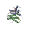
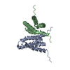
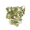
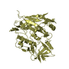
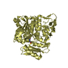
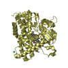
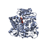
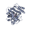
- PDB-1suy: NMR structure of the ThKaiA180C-CIIABD complex (average minimized... -
+ Open data

- Basic information
| Entry | Database: PDB / ID: 1suy | ||||||
|---|---|---|---|---|---|---|---|
| Title | NMR structure of the ThKaiA180C-CIIABD complex (average minimized structure) | ||||||
 Components Components |
| ||||||
 Keywords Keywords |  CIRCADIAN CLOCK PROTEIN / X-class Four Helix Bundle / Protein-peptide complex CIRCADIAN CLOCK PROTEIN / X-class Four Helix Bundle / Protein-peptide complex | ||||||
| Function / homology |  Function and homology information Function and homology informationprotein serine/threonine/tyrosine kinase activity / Hydrolases; Acting on acid anhydrides; Acting on acid anhydrides to facilitate cellular and subcellular movement / regulation of circadian rhythm / circadian rhythm / kinase activity / protein autophosphorylation / non-specific serine/threonine protein kinase / protein serine kinase activity / protein serine/threonine kinase activity / regulation of DNA-templated transcription ...protein serine/threonine/tyrosine kinase activity / Hydrolases; Acting on acid anhydrides; Acting on acid anhydrides to facilitate cellular and subcellular movement / regulation of circadian rhythm / circadian rhythm / kinase activity / protein autophosphorylation / non-specific serine/threonine protein kinase / protein serine kinase activity / protein serine/threonine kinase activity / regulation of DNA-templated transcription / magnesium ion binding / ATP hydrolysis activity / DNA binding / ATP binding / identical protein bindingSimilarity search - Function | ||||||
| Biological species |   Thermosynechococcus elongatus (bacteria) Thermosynechococcus elongatus (bacteria) | ||||||
| Method | SOLUTION NMR / Distance geometry, Simulated annealing | ||||||
| Model type details | minimized average | ||||||
 Authors Authors | Vakonakis, I. / LiWang, A.C. | ||||||
 Citation Citation | Journal: Proc.Natl.Acad.Sci.Usa / Year: 2004 Title: Structure of the C-terminal domain of the clock protein KaiA in complex with a KaiC-derived peptide: implications for KaiC regulation. Authors: Vakonakis, I. / LiWang, A.C. | ||||||
| History |
|
- Structure visualization
| Structure viewer | Molecule: MolmilJmol/JSmol |
|---|
- Downloads & links
-Download
| PDBx/mmCIF format | 1suy.cif.gz | 108.5 KB | Display | PDBx/mmCIF format |
|---|---|---|---|---|
| PDB format | pdb1suy.ent.gz | 90.7 KB | Display | PDB format |
| PDBx/mmJSON format | 1suy.json.gz | Tree view | PDBx/mmJSON format | |
| Others |  Other downloads Other downloads |
-Validation report
| Arichive directory | https://data.pdbj.org/pub/pdb/validation_reports/su/1suyftp://data.pdbj.org/pub/pdb/validation_reports/su/1suy | HTTPS FTP |
|---|
-Related structure data


-Links
 PDBj
PDBj

- Assembly
| Deposited unit | 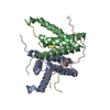
| |||||||||
|---|---|---|---|---|---|---|---|---|---|---|
| 1 |
| |||||||||
| NMR ensembles |
|
-Components
| #1: Protein | / ThKaiA180C Mass: 12601.585 Da / Num. of mol.: 2 / Fragment: C-terminal residues 180-283 Source method: isolated from a genetically manipulated source Source: (gene. exp.) Thermosynechococcus elongatus (bacteria)Strain: BP-1 / Gene: KaiA / Plasmid: pET32a+ / Species (production host): Escherichia coli / Production host: Escherichia coli BL21(DE3) (bacteria) / Strain (production host): BL21(DE3) / References: UniProt: Q79V62#2: Protein/peptide | / CIIABDMass: 3623.117 Da / Num. of mol.: 2 / Fragment: C-terminal residues 488-518 Source method: isolated from a genetically manipulated source Source: (gene. exp.) Thermosynechococcus elongatus (bacteria)Strain: BP-1 / Gene: KaiC / Plasmid: pET32a+ / Species (production host): Escherichia coli / Production host: Escherichia coli BL21(DE3) (bacteria) / Strain (production host): BL21(DE3) / References: UniProt: Q8RR33, UniProt: Q79V60*PLUS |
|---|
-Experimental details
-Experiment
| Experiment | Method: SOLUTION NMR | ||||||||||||||||||||||||
|---|---|---|---|---|---|---|---|---|---|---|---|---|---|---|---|---|---|---|---|---|---|---|---|---|---|
| NMR experiment |
| ||||||||||||||||||||||||
| NMR details | Text: Assignments of ThKaiA180C and CIIABD were performed through CBCA(CO)NH, CBCANH, HBHA(CO)NH and HC(C)H-COSY experiments |
- Sample preparation
| Details |
| |||||||||||||||
|---|---|---|---|---|---|---|---|---|---|---|---|---|---|---|---|---|
| Sample conditions | Ionic strength: 20 mM NaCl, 20 mM NaPi / pH: 7 / Pressure: ambient / Temperature: 323 K |
-NMR measurement
| Radiation | Protocol: SINGLE WAVELENGTH / Monochromatic (M) / Laue (L): M / Scattering type: x-ray | |||||||||||||||
|---|---|---|---|---|---|---|---|---|---|---|---|---|---|---|---|---|
| Radiation wavelength | Relative weight: 1 | |||||||||||||||
| NMR spectrometer |
|
- Processing
| NMR software |
| ||||||||||||||||||
|---|---|---|---|---|---|---|---|---|---|---|---|---|---|---|---|---|---|---|---|
| Refinement | Method: Distance geometry, Simulated annealing / Software ordinal: 1 Details: The structure is based on a total of 2420 restraints: 1858 are NOE-derived, 58 are from hydrogen bonds, 244 are dihedral angles and 260 are 13C chemical shifts. | ||||||||||||||||||
| NMR representative | Selection criteria: minimized average structure | ||||||||||||||||||
| NMR ensemble | Conformers submitted total number: 1 |
