 Movie
Movie Controller

[English] 日本語
 Yorodumi
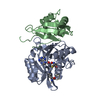
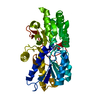
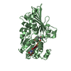
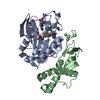
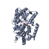
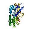
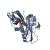
- PDB-1sqh: X-RAY STRUCTURE OF DROSOPHILA MALONOGASTER PROTEIN Q9VR51 NORTHEA... -
+ Open data

- Basic information
| Entry | Database: PDB / ID: 1sqh | ||||||
|---|---|---|---|---|---|---|---|
| Title | X-RAY STRUCTURE OF DROSOPHILA MALONOGASTER PROTEIN Q9VR51 NORTHEAST STRUCTURAL GENOMICS CONSORTIUM TARGET FR87. | ||||||
 Components Components | hypothetical protein CG14615-PA Hypothesis Hypothesis | ||||||
 Keywords Keywords | STRUCTURAL GENOMICS / UNKNOWN FUNCTION / PSI / Protein Structure Initiative / Northeast Structural Genomics Consortium / NESG | ||||||
| Function / homology |  Function and homology information Function and homology informationacyltransferase activity, transferring groups other than amino-acyl groups / Transferases; Acyltransferases; Transferring groups other than aminoacyl groupsSimilarity search - Function | ||||||
| Biological species |  Drosophila melanogaster (fruit fly) Drosophila melanogaster (fruit fly) | ||||||
| Method | X-RAY DIFFRACTION / SYNCHROTRON / SAD / Resolution: 2 Å | ||||||
 Authors Authors | Kuzin, A.P. / Chen, Y. / Forouhar, F. / Acton, T.B. / Xiao, R. / Cooper, B. / Ho, C.K. / Montelione, G.T. / Tong, L. / Hunt, J.F. / Northeast Structural Genomics Consortium (NESG) | ||||||
 Citation Citation | Journal: To be Published Title: X-RAY STRUCTURE OF DROSOPHILA MALONOGASTER PROTEIN Q9VR51 NORTHEAST STRUCTURAL GENOMICS CONSORTIUM TARGET FR87 Authors: Kuzin, A.P. / Chen, Y. / Forouhar, F. / Acton, T.B. / Xiao, R. / Cooper, B. / Ho, C.K. / Montelione, G.T. / Tong, L. / Hunt, J.F. | ||||||
| History |
|
- Structure visualization
| Structure viewer | Molecule: MolmilJmol/JSmol |
|---|
- Downloads & links
-Download
| PDBx/mmCIF format | 1sqh.cif.gz | 71.6 KB | Display | PDBx/mmCIF format |
|---|---|---|---|---|
| PDB format | pdb1sqh.ent.gz | 57.4 KB | Display | PDB format |
| PDBx/mmJSON format | 1sqh.json.gz | Tree view | PDBx/mmJSON format | |
| Others |  Other downloads Other downloads |
-Validation report
| Arichive directory | https://data.pdbj.org/pub/pdb/validation_reports/sq/1sqhftp://data.pdbj.org/pub/pdb/validation_reports/sq/1sqh | HTTPS FTP |
|---|
-Related structure data
| Similar structure data | |
|---|---|
| Other databases |


-Links
 PDBj
PDBj

- Assembly
| Deposited unit | 
| ||||||||
|---|---|---|---|---|---|---|---|---|---|
| 1 |
| ||||||||
| Unit cell |
|
-Components
| #1: Protein | Hypothesis Mass: 36137.445 Da / Num. of mol.: 1 Source method: isolated from a genetically manipulated source Source: (gene. exp.) Drosophila melanogaster (fruit fly) / Production host:  Escherichia coli (E. coli) / References: UniProt: Q9VR51 Escherichia coli (E. coli) / References: UniProt: Q9VR51 |
|---|---|
| #2: Water | ChemComp-HOH / Water Mass: 18.015 Da / Num. of mol.: 152 / Source method: isolated from a natural source / Formula: H2O Mass: 18.015 Da / Num. of mol.: 152 / Source method: isolated from a natural source / Formula: H2O |
-Experimental details
-Experiment
| Experiment | Method: X-RAY DIFFRACTION / Number of used crystals: 1 |
|---|
- Sample preparation
| Crystal | Density Matthews: 2.564 Å3/Da / Density % sol: 50.18 % |
|---|---|
| Crystal grow | Temperature: 277 K / pH: 7 Details: 100 mM ammonium sulfate, 100 mM Tris, Spermidine , pH 7.0, VAPOR DIFFUSION, HANGING DROP, temperature 277K, pH 7.00 |
-Data collection
| Diffraction | Mean temperature: 100 K |
|---|---|
| Diffraction source | Source: SYNCHROTRON / Site: NSLS  / Beamline: X4A / Wavelength: 0.97933 / Beamline: X4A / Wavelength: 0.97933 |
| Detector | Type: ADSC QUANTUM 4 / Detector: CCD / Date: Jan 31, 2004 |
| Radiation | Protocol: SINGLE WAVELENGTH / Monochromatic (M) / Laue (L): M / Scattering type: x-ray |
| Radiation wavelength | Wavelength: 0.97933 Å / Relative weight: 1 |
| Reflection | Resolution: 2→30 Å / Num. obs: 43215 / % possible obs: 88.8 % / Observed criterion σ(I): -3 / Redundancy: 3.25 % / Biso Wilson estimate: 26.3 Å2 / Rmerge(I) obs: 0.109 / Rsym value: 0.109 |
| Reflection shell | Resolution: 2→2.07 Å / % possible all: 49.7 |
- Processing
| Software |
| ||||||||||||||||||||||||||||||||||||||||||||||||||||||||||||||||||||||||||||||||
|---|---|---|---|---|---|---|---|---|---|---|---|---|---|---|---|---|---|---|---|---|---|---|---|---|---|---|---|---|---|---|---|---|---|---|---|---|---|---|---|---|---|---|---|---|---|---|---|---|---|---|---|---|---|---|---|---|---|---|---|---|---|---|---|---|---|---|---|---|---|---|---|---|---|---|---|---|---|---|---|---|---|
| Refinement | Method to determine structure: SAD / Resolution: 2→19.84 Å / Rfactor Rfree error: 0.006 / Data cutoff high absF: 346738.81 / Data cutoff low absF: 0 / Isotropic thermal model: RESTRAINED / Cross valid method: THROUGHOUT / σ(F): 2 / Stereochemistry target values: ENGH & HUBER Details: 8542 REFLECTIONS ARE UNOBSERVED (NO ENTRY OR |F|=0)
| ||||||||||||||||||||||||||||||||||||||||||||||||||||||||||||||||||||||||||||||||
| Solvent computation | Solvent model: FLAT MODEL / Bsol: 32.42 Å2 / ksol: 0.3 e/Å3 | ||||||||||||||||||||||||||||||||||||||||||||||||||||||||||||||||||||||||||||||||
| Displacement parameters | Biso mean: 31.3 Å2
| ||||||||||||||||||||||||||||||||||||||||||||||||||||||||||||||||||||||||||||||||
| Refine analyze |
| ||||||||||||||||||||||||||||||||||||||||||||||||||||||||||||||||||||||||||||||||
| Refinement step | Cycle: LAST / Resolution: 2→19.84 Å
| ||||||||||||||||||||||||||||||||||||||||||||||||||||||||||||||||||||||||||||||||
| Refine LS restraints |
| ||||||||||||||||||||||||||||||||||||||||||||||||||||||||||||||||||||||||||||||||
| LS refinement shell | Resolution: 2→2.13 Å / Rfactor Rfree error: 0.033 / Total num. of bins used: 6
| ||||||||||||||||||||||||||||||||||||||||||||||||||||||||||||||||||||||||||||||||
| Xplor file |
|
