Mass: 18.015 Da / Num. of mol.: 822 / Source method: isolated from a natural source / Formula: H2O
-
Experimental details
-
Experiment
Experiment
Method: X-RAY DIFFRACTION / Number of used crystals: 1
-
Sample preparation
Crystal
Density Matthews: 2.9 Å3/Da / Density % sol: 58 %
Crystal grow
Method: vapor diffusion, hanging drop / pH: 7.5 Details: PROTEIN WAS CRYSTALLIZED BY VAPOR DIFFUSION USING HANGING DROPS AND USING MICROSEEDING. RESERVOIR: 0.65-0.9 M LI2SO4, 100 MM HEPES PH 7.5, vapor diffusion - hanging drop
Crystal grow
*PLUS
Temperature: 22 ℃ / Method: vapor diffusion / Details: used to seeding
Monochromatic (M) / Laue (L): M / Scattering type: x-ray
Radiation wavelength
Wavelength: 1.08 Å / Relative weight: 1
Reflection
Resolution: 1.9→50 Å / Num. obs: 84147 / % possible obs: 89.2 % / Redundancy: 2.1 % / Biso Wilson estimate: 22.3 Å2 / Rsym value: 0.06 / Net I/σ(I): 11.4
Reflection shell
Resolution: 1.9→1.94 Å / Redundancy: 1.8 % / Rsym value: 0.16 / % possible all: 66.2
Reflection
*PLUS
Rmerge(I) obs: 0.06
Reflection shell
*PLUS
% possible obs: 66.2 % / Rmerge(I) obs: 0.16
-
Processing
Software
Name
Version
Classification
DENZO
datareduction
SCALEPACK
datascaling
X-PLOR
3.8
modelbuilding
X-PLOR
3.8
refinement
X-PLOR
3.8
phasing
Refinement
Method to determine structure: MIR / Resolution: 1.9→50 Å / Isotropic thermal model: RESTRAINED / Cross valid method: THROUGHOUT Details: REFINEMENT WAS CARRIED OUT USING BOTH X-PLOR AND REFMAC.
Rfactor
Num. reflection
% reflection
Selection details
Rfree
0.22
2064
2.5 %
RANDOM
Rwork
0.1748
-
-
-
obs
0.1748
84147
89.2 %
-
Displacement parameters
Biso mean: 26.1 Å2
Baniso -1
Baniso -2
Baniso -3
1-
2.641 Å2
0 Å2
0.435 Å2
2-
-
-4.339 Å2
0 Å2
3-
-
-
1.698 Å2
Refinement step
Cycle: LAST / Resolution: 1.9→50 Å
Protein
Nucleic acid
Ligand
Solvent
Total
Num. atoms
7129
0
177
822
8128
Refine LS restraints
Refine-ID
Type
Dev ideal
Dev ideal target
X-RAY DIFFRACTION
x_bond_d
0.009
X-RAY DIFFRACTION
x_bond_d_na
X-RAY DIFFRACTION
x_bond_d_prot
X-RAY DIFFRACTION
x_angle_d
X-RAY DIFFRACTION
x_angle_d_na
X-RAY DIFFRACTION
x_angle_d_prot
X-RAY DIFFRACTION
x_angle_deg
1.4
X-RAY DIFFRACTION
x_angle_deg_na
X-RAY DIFFRACTION
x_angle_deg_prot
X-RAY DIFFRACTION
x_dihedral_angle_d
X-RAY DIFFRACTION
x_dihedral_angle_d_na
X-RAY DIFFRACTION
x_dihedral_angle_d_prot
X-RAY DIFFRACTION
x_improper_angle_d
X-RAY DIFFRACTION
x_improper_angle_d_na
X-RAY DIFFRACTION
x_improper_angle_d_prot
X-RAY DIFFRACTION
x_mcbond_it
2.43
2
X-RAY DIFFRACTION
x_mcangle_it
3.22
3
X-RAY DIFFRACTION
x_scbond_it
2.76
2
X-RAY DIFFRACTION
x_scangle_it
3.9
3
LS refinement shell
Resolution: 1.9→1.99 Å / Total num. of bins used: 8
In the structure databanks used in Yorodumi, some data are registered as the other names, "COVID-19 virus" and "2019-nCoV". Here are the details of the virus and the list of structure data.
Jan 31, 2019. EMDB accession codes are about to change! (news from PDBe EMDB page)
EMDB accession codes are about to change! (news from PDBe EMDB page)
The allocation of 4 digits for EMDB accession codes will soon come to an end. Whilst these codes will remain in use, new EMDB accession codes will include an additional digit and will expand incrementally as the available range of codes is exhausted. The current 4-digit format prefixed with “EMD-” (i.e. EMD-XXXX) will advance to a 5-digit format (i.e. EMD-XXXXX), and so on. It is currently estimated that the 4-digit codes will be depleted around Spring 2019, at which point the 5-digit format will come into force.
The EM Navigator/Yorodumi systems omit the EMD- prefix.
Related info.:Q: What is EMD? / ID/Accession-code notation in Yorodumi/EM Navigator
Yorodumi is a browser for structure data from EMDB, PDB, SASBDB, etc.
This page is also the successor to EM Navigator detail page, and also detail information page/front-end page for Omokage search.
The word "yorodu" (or yorozu) is an old Japanese word meaning "ten thousand". "mi" (miru) is to see.
Related info.:EMDB / PDB / SASBDB / Comparison of 3 databanks / Yorodumi Search / Aug 31, 2016. New EM Navigator & Yorodumi / Yorodumi Papers / Jmol/JSmol / Function and homology information / Changes in new EM Navigator and Yorodumi
 Movie
Movie Controller


 Open data
 Basic information
 Components
 Keywords
 Function and homology information
 Authors
 Citation
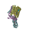
 Structure visualization
 Downloads & links
 Other downloads
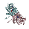


 PDBj
PDBj

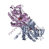
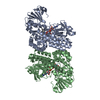
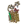
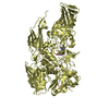
 Assembly
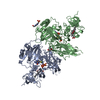


 Mass: 96.063 Da / Num. of mol.: 4 / Source method: obtained synthetically / Formula: SO4
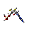
 Mass: 395.352 Da / Num. of mol.: 2 / Source method: obtained synthetically / Formula: C10H14N5O6PS2
 Mass: 95.940 Da / Num. of mol.: 2 / Source method: obtained synthetically / Formula: Mo
 Mass: 616.487 Da / Num. of mol.: 2 / Source method: obtained synthetically / Formula: C34H32FeN4O4
 Mass: 92.094 Da / Num. of mol.: 1 / Source method: obtained synthetically / Formula: C3H8O3
 Mass: 238.305 Da / Num. of mol.: 1 / Source method: obtained synthetically / Formula: C8H18N2O4S / Comment: pH buffer*YM
 Sample preparation
 / Beamline: BL7-1 / Wavelength: 1.08
  Processing