 Movie
Movie Controller


+ Open data

- Basic information
| Entry | Database: PDB / ID: 1sny | ||||||
|---|---|---|---|---|---|---|---|
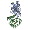
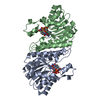
| Title | Carbonyl reductase Sniffer of D. melanogaster | ||||||
 Components Components | sniffer CG10964-PA | ||||||
 Keywords Keywords |  OXIDOREDUCTASE / alpha and beta protein / Rossmann fold / dinucleotide binding motif OXIDOREDUCTASE / alpha and beta protein / Rossmann fold / dinucleotide binding motif | ||||||
| Function / homology |  Function and homology informationcarbonyl reductase (NADPH) / carbonyl reductase (NADPH) activity / : / nucleotide binding Function and homology informationcarbonyl reductase (NADPH) / carbonyl reductase (NADPH) activity / : / nucleotide bindingSimilarity search - Function | ||||||
| Biological species |  Drosophila melanogaster (fruit fly) Drosophila melanogaster (fruit fly) | ||||||
| Method | X-RAY DIFFRACTION / SYNCHROTRON / MAD / Resolution: 1.75 Å | ||||||
 Authors Authors | Sgraja, T. / Ulschmid, J. / Becker, K. / Schneuwly, S. / Klebe, G. / Reuter, K. / Heine, A. | ||||||
 Citation Citation | Journal: J.Mol.Biol. / Year: 2004 Title: Structural Insights into the Neuroprotective-acting Carbonyl Reductase Sniffer of Drosophila melanogaster. Authors: Sgraja, T. / Ulschmid, J. / Becker, K. / Schneuwly, S. / Klebe, G. / Reuter, K. / Heine, A. | ||||||
| History |
|
- Structure visualization
| Structure viewer | Molecule: MolmilJmol/JSmol |
|---|
- Downloads & links
-Download
| PDBx/mmCIF format | 1sny.cif.gz | 58.1 KB | Display | PDBx/mmCIF format |
|---|---|---|---|---|
| PDB format | pdb1sny.ent.gz | 45.9 KB | Display | PDB format |
| PDBx/mmJSON format | 1sny.json.gz | Tree view | PDBx/mmJSON format | |
| Others |  Other downloads Other downloads |
-Validation report
| Arichive directory | https://data.pdbj.org/pub/pdb/validation_reports/sn/1snyftp://data.pdbj.org/pub/pdb/validation_reports/sn/1sny | HTTPS FTP |
|---|
-Related structure data
| Similar structure data |
|---|


-Links
 PDBj
PDBj

- Assembly
| Deposited unit | 
| ||||||||
|---|---|---|---|---|---|---|---|---|---|
| 1 | 
| ||||||||
| Unit cell |
|
-Components
| #1: Protein | Mass: 29040.141 Da / Num. of mol.: 1 Source method: isolated from a genetically manipulated source Source: (gene. exp.) Drosophila melanogaster (fruit fly) / Gene: sniffer / Plasmid: pQE16 / Production host:  Escherichia coli (E. coli) / Strain (production host): M15 (Qiagen) / References: UniProt: Q9W3H4 Escherichia coli (E. coli) / Strain (production host): M15 (Qiagen) / References: UniProt: Q9W3H4 |
|---|---|
| #2: Chemical | ChemComp-NAP / Nicotinamide adenine dinucleotide phosphate  Mass: 743.405 Da / Num. of mol.: 1 / Source method: obtained synthetically / Formula: C21H28N7O17P3 Mass: 743.405 Da / Num. of mol.: 1 / Source method: obtained synthetically / Formula: C21H28N7O17P3 |
| #3: Water | ChemComp-HOH / Water Mass: 18.015 Da / Num. of mol.: 57 / Source method: isolated from a natural source / Formula: H2O Mass: 18.015 Da / Num. of mol.: 57 / Source method: isolated from a natural source / Formula: H2O |
-Experimental details
-Experiment
| Experiment | Method: X-RAY DIFFRACTION / Number of used crystals: 1 |
|---|
- Sample preparation
| Crystal | Density Matthews: 2.11 Å3/Da / Density % sol: 41.57 % |
|---|---|
| Crystal grow | Temperature: 277 K / Method: vapor diffusion, hanging drop / pH: 8.5 Details: PEG 4000, sodium acetate, tris hydrogen chloride, pH 8.5, VAPOR DIFFUSION, HANGING DROP, temperature 277K |
-Data collection
| Diffraction | Mean temperature: 100 K | ||||||||||||
|---|---|---|---|---|---|---|---|---|---|---|---|---|---|
| Diffraction source | Source: SYNCHROTRON / Site: BESSY  / Beamline: 14.2 / Wavelength: 0.980163, 0.979944, 0.932328 / Beamline: 14.2 / Wavelength: 0.980163, 0.979944, 0.932328 | ||||||||||||
| Detector | Type: MAR scanner 345 mm plate / Detector: IMAGE PLATE / Date: May 22, 2003 / Details: mirrors | ||||||||||||
| Radiation | Monochromator: Si-111 crystal / Protocol: MAD / Monochromatic (M) / Laue (L): M / Scattering type: x-ray | ||||||||||||
| Radiation wavelength |
| ||||||||||||
| Reflection | Resolution: 1.75→28.84 Å / Num. obs: 24381 / % possible obs: 97.5 % / Observed criterion σ(F): -3 / Observed criterion σ(I): -3 / Redundancy: 7.3 % / Biso Wilson estimate: 19.3 Å2 / Rmerge(I) obs: 0.073 / Rsym value: 0.073 / Net I/σ(I): 22.7 | ||||||||||||
| Reflection shell | Resolution: 1.75→1.86 Å / Redundancy: 7.3 % / Rmerge(I) obs: 0.498 / Mean I/σ(I) obs: 3.1 / Num. unique all: 24381 / Rsym value: 0.498 / % possible all: 98.7 |
- Processing
| Software |
| ||||||||||||||||||||||||||||||||||||
|---|---|---|---|---|---|---|---|---|---|---|---|---|---|---|---|---|---|---|---|---|---|---|---|---|---|---|---|---|---|---|---|---|---|---|---|---|---|
| Refinement | Method to determine structure: MAD / Resolution: 1.75→28.84 Å / Rfactor Rfree error: 0.009 / Data cutoff high absF: 1209442.33 / Data cutoff low absF: 0 / Isotropic thermal model: anisotropic / Cross valid method: THROUGHOUT / σ(F): 0 / Stereochemistry target values: Engh & Huber Details: poorly defined sections in the structure: amino acid 10 to 18, 22 to 26 and 37 to 53
| ||||||||||||||||||||||||||||||||||||
| Solvent computation | Solvent model: FLAT MODEL / Bsol: 48.8512 Å2 / ksol: 0.376692 e/Å3 | ||||||||||||||||||||||||||||||||||||
| Displacement parameters | Biso mean: 33.7 Å2
| ||||||||||||||||||||||||||||||||||||
| Refine analyze |
| ||||||||||||||||||||||||||||||||||||
| Refinement step | Cycle: LAST / Resolution: 1.75→28.84 Å
| ||||||||||||||||||||||||||||||||||||
| Refine LS restraints |
| ||||||||||||||||||||||||||||||||||||
| LS refinement shell | Resolution: 1.75→1.86 Å / Rfactor Rfree error: 0.037 / Total num. of bins used: 6
| ||||||||||||||||||||||||||||||||||||
| Xplor file |
|
