 Movie
Movie Controller


+ Open data

- Basic information
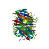
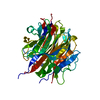
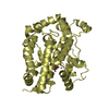
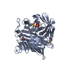
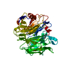
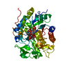
| Entry | Database: PDB / ID: 1snt | ||||||
|---|---|---|---|---|---|---|---|
| Title | Structure of the human cytosolic sialidase Neu2 | ||||||
 Components Components | Sialidase 2 Neuraminidase Neuraminidase | ||||||
 Keywords Keywords | HYDROLASE / sialidase / neuraminidase / ganglioside | ||||||
| Function / homology |  Function and homology information Function and homology informationSialic acid metabolism / glycoprotein catabolic process / glycosphingolipid catabolic process / ganglioside catabolic process / exo-alpha-sialidase activity / oligosaccharide catabolic process / Glycosphingolipid catabolism / exo-alpha-(2->3)-sialidase activity / exo-alpha-(2->6)-sialidase activity / exo-alpha-(2->8)-sialidase activity ...Sialic acid metabolism / glycoprotein catabolic process / glycosphingolipid catabolic process / ganglioside catabolic process / exo-alpha-sialidase activity / oligosaccharide catabolic process / Glycosphingolipid catabolism / exo-alpha-(2->3)-sialidase activity / exo-alpha-(2->6)-sialidase activity / exo-alpha-(2->8)-sialidase activity / exo-alpha-sialidase / catalytic complex / lysosome / membrane / cytosol / cytoplasmSimilarity search - Function | ||||||
| Biological species |  Homo sapiens (human) Homo sapiens (human) | ||||||
| Method | X-RAY DIFFRACTION / SYNCHROTRON / MOLECULAR REPLACEMENT / Resolution: 1.75 Å | ||||||
 Authors Authors | Chavas, L.M.G. / Fusi, P. / Tringali, C. / Venerando, B. / Tettamanti, G. / Kato, R. / Monti, E. / Wakatsuki, S. | ||||||
 Citation Citation | Journal: J.Biol.Chem. / Year: 2005 Title: Crystal Structure of the Human Cytosolic Sialidase Neu2: EVIDENCE FOR THE DYNAMIC NATURE OF SUBSTRATE RECOGNITION Authors: Chavas, L.M.G. / Tringali, C. / Fusi, P. / Venerando, B. / Tettamanti, G. / Kato, R. / Monti, E. / Wakatsuki, S. | ||||||
| History |
|
- Structure visualization
| Structure viewer | Molecule: MolmilJmol/JSmol |
|---|
- Downloads & links
-Download
| PDBx/mmCIF format | 1snt.cif.gz | 92.1 KB | Display | PDBx/mmCIF format |
|---|---|---|---|---|
| PDB format | pdb1snt.ent.gz | 68.1 KB | Display | PDB format |
| PDBx/mmJSON format | 1snt.json.gz | Tree view | PDBx/mmJSON format | |
| Others |  Other downloads Other downloads |
-Validation report
| Arichive directory | https://data.pdbj.org/pub/pdb/validation_reports/sn/1sntftp://data.pdbj.org/pub/pdb/validation_reports/sn/1snt | HTTPS FTP |
|---|
-Related structure data
| Related structure data |  1so7C  1vcuC  1eusS C: citing same article ( S: Starting model for refinement |
|---|---|
| Similar structure data |


-Links
 PDBj
PDBj

- Assembly
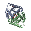
| Deposited unit | 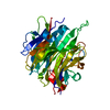
| ||||||||
|---|---|---|---|---|---|---|---|---|---|
| 1 |
| ||||||||
| Unit cell |
|
-Components
| #1: Protein | Neuraminidase / Neu2 / Cytosolic sialidase / N-acetyl-alpha- neuraminidase 2 Mass: 42419.598 Da / Num. of mol.: 1 Source method: isolated from a genetically manipulated source Source: (gene. exp.) Homo sapiens (human) / Plasmid: pGEX-2T / Species (production host): Escherichia coli / Production host:  Escherichia coli BL21 (bacteria) / Strain (production host): BL21 / References: UniProt: Q9Y3R4, exo-alpha-sialidase Escherichia coli BL21 (bacteria) / Strain (production host): BL21 / References: UniProt: Q9Y3R4, exo-alpha-sialidase |
|---|---|
| #2: Water | ChemComp-HOH / Water Mass: 18.015 Da / Num. of mol.: 377 / Source method: isolated from a natural source / Formula: H2O Mass: 18.015 Da / Num. of mol.: 377 / Source method: isolated from a natural source / Formula: H2O |
-Experimental details
-Experiment
| Experiment | Method: X-RAY DIFFRACTION / Number of used crystals: 1 |
|---|
- Sample preparation
| Crystal | Density Matthews: 2.98 Å3/Da / Density % sol: 58.35 % |
|---|---|
| Crystal grow | Temperature: 289 K / Method: vapor diffusion, hanging drop / pH: 6.2 Details: sodium potassium phosphate, sodium chloride, pH 6.2, VAPOR DIFFUSION, HANGING DROP, temperature 289K |
-Data collection
| Diffraction | Mean temperature: 100 K |
|---|---|
| Diffraction source | Source: SYNCHROTRON / Site: Photon Factory  / Beamline: AR-NW12A / Wavelength: 0.978 Å / Beamline: AR-NW12A / Wavelength: 0.978 Å |
| Detector | Type: ADSC QUANTUM 210 / Detector: CCD / Date: May 24, 2003 |
| Radiation | Monochromator: Si(111) / Protocol: SINGLE WAVELENGTH / Monochromatic (M) / Laue (L): M / Scattering type: x-ray |
| Radiation wavelength | Wavelength: 0.978 Å / Relative weight: 1 |
| Reflection | Resolution: 1.75→40 Å / Num. all: 51301 / Num. obs: 51297 / % possible obs: 99.5 % / Rmerge(I) obs: 0.072 / Net I/σ(I): 5.5 |
| Reflection shell | Resolution: 1.75→1.84 Å / Rmerge(I) obs: 0.383 / Mean I/σ(I) obs: 2 / % possible all: 99.5 |
- Processing
| Software |
| |||||||||||||||||||||||||
|---|---|---|---|---|---|---|---|---|---|---|---|---|---|---|---|---|---|---|---|---|---|---|---|---|---|---|
| Refinement | Method to determine structure: MOLECULAR REPLACEMENT Starting model: PDB ENTRY 1EUS Resolution: 1.75→40 Å / Cross valid method: THROUGHOUT / σ(F): 0 / Stereochemistry target values: Engh & Huber Details: About CD1/CD2 (LEU A 90) and CG1/CG2 (VAL A 325), there are 3 possible positions for these atoms. There are two different conformations for each atom. For convenience, the author assigned as ...Details: About CD1/CD2 (LEU A 90) and CG1/CG2 (VAL A 325), there are 3 possible positions for these atoms. There are two different conformations for each atom. For convenience, the author assigned as a double conformation only one of the atoms.(CD2 of LEU 90 and CG1 of VAL 325) Thus the occupancies of the alternate conformations are greater than 1.00 and there are chirality errors at these atoms.
| |||||||||||||||||||||||||
| Refinement step | Cycle: LAST / Resolution: 1.75→40 Å
| |||||||||||||||||||||||||
| Refine LS restraints |
|
