 Movie
Movie Controller

[English] 日本語
 Yorodumi
- PDB-1snn: 3,4-dihydroxy-2-butanone 4-phosphate synthase from Methanococcus ... -
+ Open data

- Basic information
| Entry | Database: PDB / ID: 1snn | ||||||
|---|---|---|---|---|---|---|---|
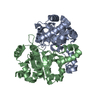
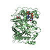
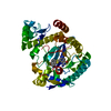
| Title | 3,4-dihydroxy-2-butanone 4-phosphate synthase from Methanococcus jannaschii | ||||||
 Components Components | 3,4-dihydroxy-2-butanone 4-phosphate synthase | ||||||
 Keywords Keywords |  ISOMERASE / riboflavin biosynthesis ISOMERASE / riboflavin biosynthesis | ||||||
| Function / homology |  Function and homology information3,4-dihydroxy-2-butanone-4-phosphate synthase / 3,4-dihydroxy-2-butanone-4-phosphate synthase activity / riboflavin biosynthetic process / manganese ion binding / magnesium ion binding / cytosol Function and homology information3,4-dihydroxy-2-butanone-4-phosphate synthase / 3,4-dihydroxy-2-butanone-4-phosphate synthase activity / riboflavin biosynthetic process / manganese ion binding / magnesium ion binding / cytosolSimilarity search - Function | ||||||
| Biological species |   Methanocaldococcus jannaschii (archaea) Methanocaldococcus jannaschii (archaea) | ||||||
| Method | X-RAY DIFFRACTION / SYNCHROTRON / FOURIER SYNTHESIS / Resolution: 1.55 Å | ||||||
 Authors Authors | Steinbacher, S. / Schiffmann, S. / Huber, R. / Bacher, A. / Fischer, M. | ||||||
 Citation Citation | Journal: Acta Crystallogr.,Sect.D / Year: 2004 Title: Metal sites in 3,4-dihydroxy-2-butanone 4-phosphate synthase from Methanococcus jannaschii in complex with the substrate ribulose 5-phosphate. Authors: Steinbacher, S. / Schiffmann, S. / Bacher, A. / Fischer, M. #1: Journal: J.Biol.Chem. / Year: 2003Title: Structure of 3,4-dihydroxy-2-butanone 4-phosphate synthase from Methanococcus jannaschii in complex with divalent metal ions and the substrate ribulose 5-phosphate: implications for the catalytic mechanism Authors: Steinbacher, S. / Schiffmann, S. / Richter, G. / Huber, R. / Bacher, A. / Fischer, M. | ||||||
| History |
|
- Structure visualization
| Structure viewer | Molecule: MolmilJmol/JSmol |
|---|
- Downloads & links
-Download
| PDBx/mmCIF format | 1snn.cif.gz | 110.5 KB | Display | PDBx/mmCIF format |
|---|---|---|---|---|
| PDB format | pdb1snn.ent.gz | 83.6 KB | Display | PDB format |
| PDBx/mmJSON format | 1snn.json.gz | Tree view | PDBx/mmJSON format | |
| Others |  Other downloads Other downloads |
-Validation report
| Arichive directory | https://data.pdbj.org/pub/pdb/validation_reports/sn/1snnftp://data.pdbj.org/pub/pdb/validation_reports/sn/1snn | HTTPS FTP |
|---|
-Related structure data
| Related structure data |  1pvyS S: Starting model for refinement |
|---|---|
| Similar structure data |


-Links
 PDBj
PDBj- Assembly
| Deposited unit | 
| ||||||||
|---|---|---|---|---|---|---|---|---|---|
| 1 |
| ||||||||
| Unit cell |
| ||||||||
| Details | The biological unit is identical to the assymmetric dimer |
-Components
| #1: Protein | Mass: 25781.553 Da / Num. of mol.: 2 / Mutation: H147S Source method: isolated from a genetically manipulated source Source: (gene. exp.) Methanocaldococcus jannaschii (archaea)Gene: MJ0055 / Plasmid: PNCO-MJ / Production host:  Escherichia coli (E. coli) / Strain (production host): XL1 Escherichia coli (E. coli) / Strain (production host): XL1References: UniProt: Q60364, Isomerases; Intramolecular transferases; Transferring other groups#2: Sugar | Ribulose 5-phosphate  Type: saccharideCarbohydrate / Mass: 230.110 Da / Num. of mol.: 2 Type: saccharideCarbohydrate / Mass: 230.110 Da / Num. of mol.: 2Source method: isolated from a genetically manipulated source Formula: C5H11O8P #3: Chemical |   Mass: 65.409 Da / Num. of mol.: 3 / Source method: obtained synthetically / Formula: Zn Mass: 65.409 Da / Num. of mol.: 3 / Source method: obtained synthetically / Formula: Zn#4: Chemical | ChemComp-CA /   Mass: 40.078 Da / Num. of mol.: 4 / Source method: obtained synthetically / Formula: Ca Mass: 40.078 Da / Num. of mol.: 4 / Source method: obtained synthetically / Formula: Ca#5: Water | ChemComp-HOH / | Water Mass: 18.015 Da / Num. of mol.: 352 / Source method: isolated from a natural source / Formula: H2O Mass: 18.015 Da / Num. of mol.: 352 / Source method: isolated from a natural source / Formula: H2O |
|---|
-Experimental details
-Experiment
| Experiment | Method: X-RAY DIFFRACTION / Number of used crystals: 1 |
|---|
- Sample preparation
| Crystal | Density Matthews: 1.97 Å3/Da / Density % sol: 37.1 % |
|---|---|
| Crystal grow | Temperature: 293 K / Method: vapor diffusion, sitting drop / pH: 7.5 Details: PEG1000, pH 7.5, VAPOR DIFFUSION, SITTING DROP, temperature 293K |
-Data collection
| Diffraction | Mean temperature: 100 K | |||||||||
|---|---|---|---|---|---|---|---|---|---|---|
| Diffraction source | Source: SYNCHROTRON / Site: MPG/DESY, HAMBURG  / Beamline: BW6 / Wavelength: 1.283, 1.2823 / Beamline: BW6 / Wavelength: 1.283, 1.2823 | |||||||||
| Detector | Type: MARRESEARCH / Detector: IMAGE PLATE / Date: Aug 15, 1999 | |||||||||
| Radiation | Protocol: SINGLE WAVELENGTH / Monochromatic (M) / Laue (L): M / Scattering type: x-ray | |||||||||
| Radiation wavelength |
| |||||||||
| Reflection | Resolution: 1.55→20 Å / Num. all: 53846 / Num. obs: 53846 / % possible obs: 0.883 % / Observed criterion σ(F): 0 / Observed criterion σ(I): 0 / Redundancy: 2.4 % / Rmerge(I) obs: 0.044 / Net I/σ(I): 17.5 | |||||||||
| Reflection shell | Resolution: 1.55→1.59 Å / Redundancy: 2.4 % / Rmerge(I) obs: 0.267 / Mean I/σ(I) obs: 2 / % possible all: 92.8 |
- Processing
| Software |
| |||||||||||||||||||||||||
|---|---|---|---|---|---|---|---|---|---|---|---|---|---|---|---|---|---|---|---|---|---|---|---|---|---|---|
| Refinement | Method to determine structure: FOURIER SYNTHESIS Starting model: 1PVY Resolution: 1.55→20 Å / Isotropic thermal model: anisotropic / Cross valid method: THROUGHOUT / σ(F): 0 / σ(I): 0 / Stereochemistry target values: Engh & Huber Details: The purpose of the present study was to unumbiguously identify a metal ion as zinc. Therefore a data set including the anomlaous data was collected at the zinc absorption edge. The structure ...Details: The purpose of the present study was to unumbiguously identify a metal ion as zinc. Therefore a data set including the anomlaous data was collected at the zinc absorption edge. The structure is virtually identical to the entry 1PVY. Therefore, a simple refinement was sufficient to "solve" the structure. The strcuture was refined against the data set, not containing the anomalous data.
| |||||||||||||||||||||||||
| Displacement parameters |
| |||||||||||||||||||||||||
| Refinement step | Cycle: LAST / Resolution: 1.55→20 Å
| |||||||||||||||||||||||||
| Refine LS restraints |
|
