 Movie
Movie Controller

[English] 日本語
 Yorodumi
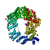
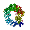
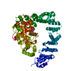
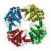
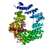
- PDB-1sly: COMPLEX OF THE 70-KDA SOLUBLE LYTIC TRANSGLYCOSYLASE WITH BULGECIN A -
+ Open data

- Basic information
| Entry | Database: PDB / ID: 1sly | ||||||
|---|---|---|---|---|---|---|---|
| Title | COMPLEX OF THE 70-KDA SOLUBLE LYTIC TRANSGLYCOSYLASE WITH BULGECIN A | ||||||
 Components Components | 70-KDA SOLUBLE LYTIC TRANSGLYCOSYLASE | ||||||
 Keywords Keywords |  GLYCOSYLTRANSFERASE / INHIBITOR-ENZYME COMPLEX / LYSOZYME / PEPTIDOGLYCAN / HYDROLASE / GLYCOSIDASE / PERIPLASMIC GLYCOSYLTRANSFERASE / INHIBITOR-ENZYME COMPLEX / LYSOZYME / PEPTIDOGLYCAN / HYDROLASE / GLYCOSIDASE / PERIPLASMIC | ||||||
| Function / homology |  Function and homology information Function and homology informationlytic endotransglycosylase activity / : / lytic transglycosylase activity / peptidoglycan metabolic process / hydrolase activity, hydrolyzing O-glycosyl compounds / peptidoglycan catabolic process / peptidoglycan-based cell wall / cell wall organization / outer membrane-bounded periplasmic space / periplasmic space ...lytic endotransglycosylase activity / : / lytic transglycosylase activity / peptidoglycan metabolic process / hydrolase activity, hydrolyzing O-glycosyl compounds / peptidoglycan catabolic process / peptidoglycan-based cell wall / cell wall organization / outer membrane-bounded periplasmic space / periplasmic space / lyase activity / membraneSimilarity search - Function | ||||||
| Biological species |  Escherichia coli (E. coli) Escherichia coli (E. coli) | ||||||
| Method | X-RAY DIFFRACTION / SYNCHROTRON / Resolution: 2.8 Å | ||||||
 Authors Authors | Thunnissen, A.M.W.H. / Kalk, K.H. / Rozeboom, H.J. / Dijkstra, B.W. | ||||||
 Citation Citation | Journal: Biochemistry / Year: 1995 Title: Structure of the 70-kDa soluble lytic transglycosylase complexed with bulgecin A. Implications for the enzymatic mechanism. Authors: Thunnissen, A.M. / Rozeboom, H.J. / Kalk, K.H. / Dijkstra, B.W. #1: Journal: Proteins / Year: 1995Title: The Catalytic Domain of a Bacterial Lytic Transglycosylase Defines a Novel Class of Lysozymes Authors: Thunnissen, A.M. / Isaacs, N.W. / Dijkstra, B.W. #2: Journal: Curr.Opin.Struct.Biol. / Year: 1994Title: 'Holy' Proteins. II: The Soluble Lytic Transglycosylase Authors: Dijkstra, B.W. / Thunnissen, A.M. #3: Journal: Nature / Year: 1994Title: Doughnut-Shaped Structure of a Bacterial Muramidase Revealed by X-Ray Crystallography Authors: Thunnissen, A.M. / Dijkstra, A.J. / Kalk, K.H. / Rozeboom, H.J. / Engel, H. / Keck, W. / Dijkstra, B.W. | ||||||
| History |
|
- Structure visualization
| Structure viewer | Molecule: MolmilJmol/JSmol |
|---|
- Downloads & links
-Download
| PDBx/mmCIF format | 1sly.cif.gz | 128.5 KB | Display | PDBx/mmCIF format |
|---|---|---|---|---|
| PDB format | pdb1sly.ent.gz | 105.5 KB | Display | PDB format |
| PDBx/mmJSON format | 1sly.json.gz | Tree view | PDBx/mmJSON format | |
| Others |  Other downloads Other downloads |
-Validation report
| Arichive directory | https://data.pdbj.org/pub/pdb/validation_reports/sl/1slyftp://data.pdbj.org/pub/pdb/validation_reports/sl/1sly | HTTPS FTP |
|---|
-Related structure data
| Similar structure data |
|---|

-Links
 PDBj
PDBj
- Assembly
| Deposited unit | 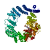
| ||||||||
|---|---|---|---|---|---|---|---|---|---|
| 1 |
| ||||||||
| Unit cell |
|
-Components
| #1: Protein | Mass: 70560.555 Da / Num. of mol.: 1 / Source method: isolated from a natural source / Source: (natural) Escherichia coli (E. coli)References: UniProt: P03810, UniProt: P0AGC3*PLUS, Hydrolases; Glycosylases; Glycosidases, i.e. enzymes that hydrolyse O- and S-glycosyl compounds |
|---|---|
| #2: Chemical | ChemComp-BLG /   Mass: 552.551 Da / Num. of mol.: 1 / Source method: obtained synthetically / Formula: C16H30N3O14S2 Mass: 552.551 Da / Num. of mol.: 1 / Source method: obtained synthetically / Formula: C16H30N3O14S2 |
| Compound details | THE STRUCTURE CONSISTS OF 3 DOMAINS: DOMAIN RESIDUES COMMENT U 1 - 361 ALPHA-SUPERHELIX TOPOLOGY L ...THE STRUCTURE CONSISTS OF 3 DOMAINS: DOMAIN RESIDUES COMMENT U 1 - 361 ALPHA-SUPERHELIX |
-Experimental details
-Experiment
| Experiment | Method: X-RAY DIFFRACTION |
|---|
- Sample preparation
| Crystal | Density Matthews: 3.35 Å3/Da / Density % sol: 63.2 % | |||||||||||||||||||||||||
|---|---|---|---|---|---|---|---|---|---|---|---|---|---|---|---|---|---|---|---|---|---|---|---|---|---|---|
| Crystal | *PLUS Density % sol: 63 % | |||||||||||||||||||||||||
| Crystal grow | *PLUS pH: 6.8 / Method: vapor diffusion, hanging drop / Details: Rozeboom, H. J., (1990) J. Mol. Biol., 212, 557. | |||||||||||||||||||||||||
| Components of the solutions | *PLUS
|
-Data collection
| Diffraction source | Source: SYNCHROTRON / Site: EMBL/DESY, Hamburg  / Beamline: X11 / Wavelength: 1.003 / Beamline: X11 / Wavelength: 1.003 |
|---|---|
| Detector | Type: MARRESEARCH / Detector: IMAGE PLATE / Date: Oct 20, 1993 |
| Radiation | Monochromatic (M) / Laue (L): M / Scattering type: x-ray |
| Radiation wavelength | Wavelength: 1.003 Å / Relative weight: 1 |
| Reflection | Resolution: 2.8→22 Å / Num. obs: 23903 / % possible obs: 99 % / Observed criterion σ(I): 0 / Redundancy: 4.8 % / Rmerge(I) obs: 0.08 |
- Processing
| Software |
| ||||||||||||||||||||||||||||||||||||||||||||||||||||||||||||||||||||||||||||||||
|---|---|---|---|---|---|---|---|---|---|---|---|---|---|---|---|---|---|---|---|---|---|---|---|---|---|---|---|---|---|---|---|---|---|---|---|---|---|---|---|---|---|---|---|---|---|---|---|---|---|---|---|---|---|---|---|---|---|---|---|---|---|---|---|---|---|---|---|---|---|---|---|---|---|---|---|---|---|---|---|---|---|
| Refinement | Resolution: 2.8→8 Å / σ(F): 2 Details: RESIDUES 371 - 377 ARE POORLY DEFINED BY THE ELECTRON DENSITY. THESE RESIDUES BELONG TO A LONG SURFACE LOOP THAT CONNECTS THE U- AND L-DOMAINS.
| ||||||||||||||||||||||||||||||||||||||||||||||||||||||||||||||||||||||||||||||||
| Displacement parameters | Biso mean: 24.15 Å2 | ||||||||||||||||||||||||||||||||||||||||||||||||||||||||||||||||||||||||||||||||
| Refine analyze | Luzzati coordinate error obs: 0.35 Å / Luzzati sigma a obs: 0.4 Å | ||||||||||||||||||||||||||||||||||||||||||||||||||||||||||||||||||||||||||||||||
| Refinement step | Cycle: LAST / Resolution: 2.8→8 Å
| ||||||||||||||||||||||||||||||||||||||||||||||||||||||||||||||||||||||||||||||||
| Refine LS restraints |
| ||||||||||||||||||||||||||||||||||||||||||||||||||||||||||||||||||||||||||||||||
| Xplor file |
| ||||||||||||||||||||||||||||||||||||||||||||||||||||||||||||||||||||||||||||||||
| Software | *PLUS Name: X-PLOR / Classification: refinement | ||||||||||||||||||||||||||||||||||||||||||||||||||||||||||||||||||||||||||||||||
| Refinement | *PLUS Highest resolution: 2.8 Å / Num. reflection obs: 22746 / Rfactor obs: 0.195 | ||||||||||||||||||||||||||||||||||||||||||||||||||||||||||||||||||||||||||||||||
| Solvent computation | *PLUS | ||||||||||||||||||||||||||||||||||||||||||||||||||||||||||||||||||||||||||||||||
| Displacement parameters | *PLUS | ||||||||||||||||||||||||||||||||||||||||||||||||||||||||||||||||||||||||||||||||
| Refine LS restraints | *PLUS
|
